. 2018 Jul 26:8:254.
doi: 10.3389/fcimb.2018.00254. eCollection 2018.

The Human Centrosomal Protein CCDC146 Binds Chlamydia trachomatis Inclusion Membrane Protein CT288 and Is Recruited to the Periphery of the Chlamydia-Containing Vacuole

Affiliations

The Human Centrosomal Protein CCDC146 Binds Chlamydia trachomatis Inclusion Membrane Protein CT288 and Is Recruited to the Periphery of the Chlamydia-Containing Vacuole

Filipe Almeida et al. Front Cell Infect Microbiol. .

Abstract

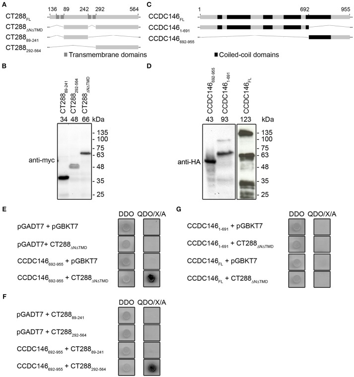
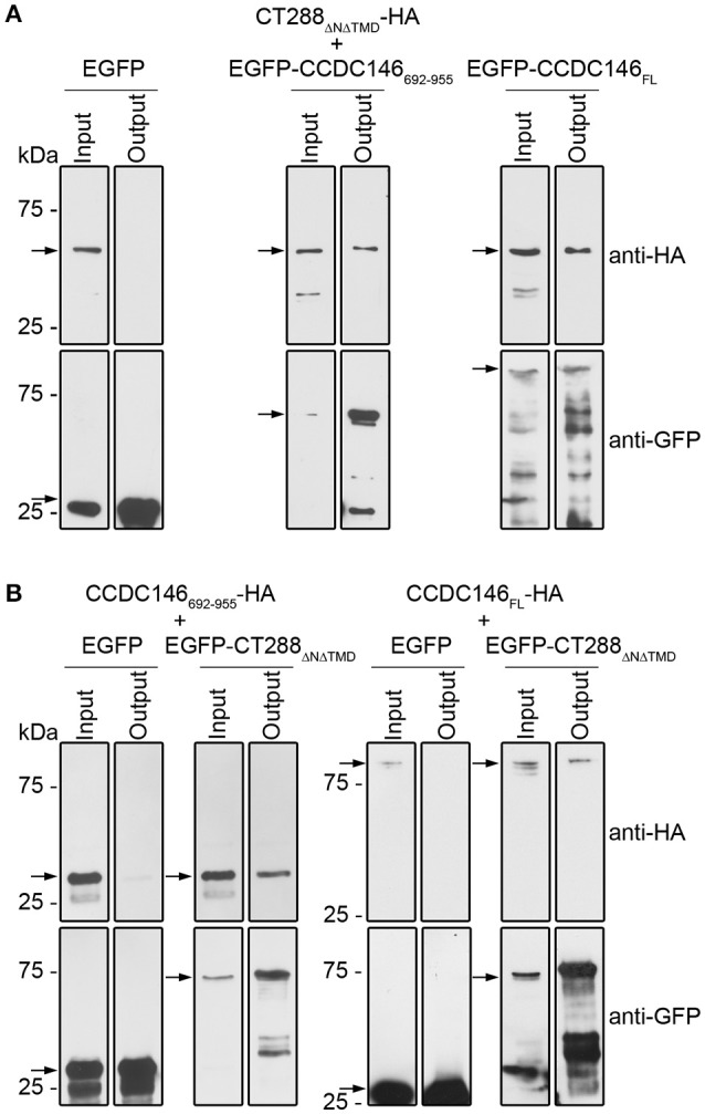
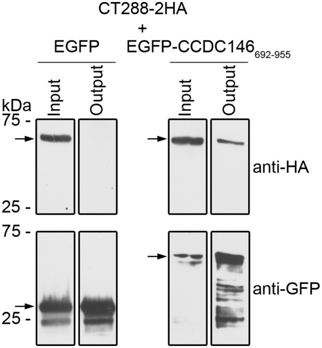
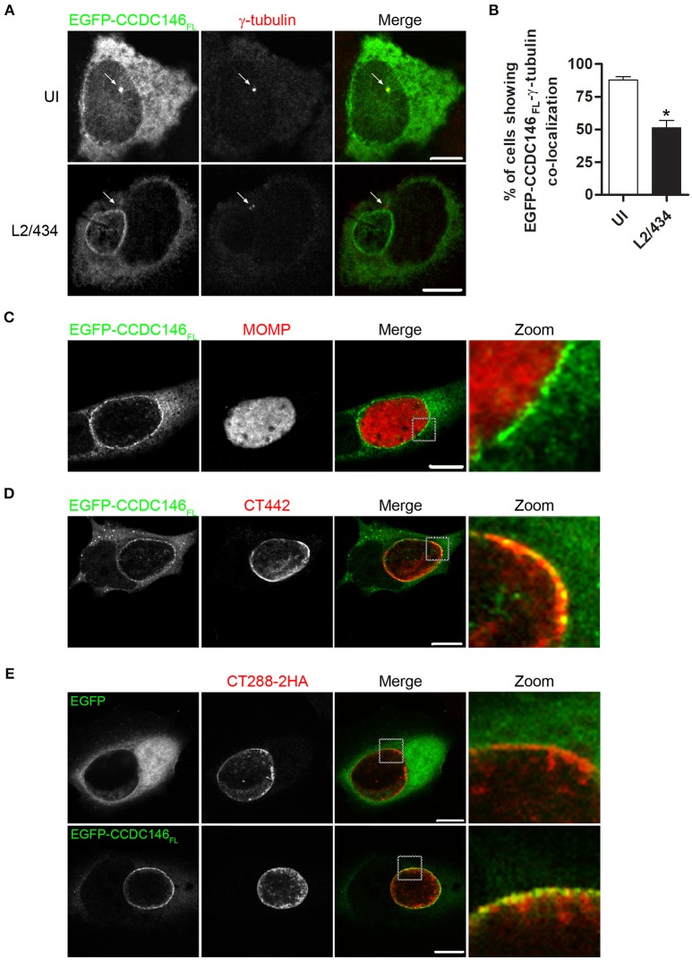
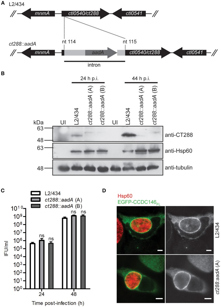
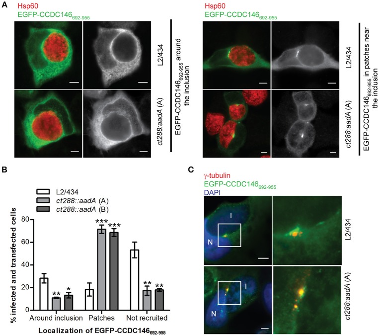
Chlamydia trachomatis is an obligate intracellular human pathogen causing mainly ocular and genital infections of significant clinical and public health impact. C. trachomatis multiplies intracellularly in a membrane bound vacuole, known as inclusion. Both extracellularly and from within the inclusion, C. trachomatis uses a type III secretion system to deliver several effector proteins into the cytoplasm of host cells. A large proportion of these effectors, the inclusion membrane (Inc) proteins, are exposed to the host cell cytosol but possess a characteristic hydrophobic domain mediating their insertion in the inclusion membrane. By yeast two-hybrid, we found that C. trachomatis Inc CT288 interacts with the human centrosomal protein CCDC146 (coiled-coil domain-containing protein 146). The interaction was also detected by co-immunoprecipitation in mammalian cells either ectopically expressing CCDC146 and CT288 or ectopically expressing CCDC146 and infected by a C. trachomatis strain expressing epitope-tagged and inclusion membrane-localized CT288. In uninfected mammalian cells, ectopically expressed full-length CCDC146 (955 amino acid residues) localized at the centrosome; but in cells infected by wild-type C. trachomatis, its centrosomal localization was less evident and CCDC146 accumulated around the inclusion. Recruitment of CCDC146 to the inclusion periphery did not require intact host Golgi, microtubules or microfilaments, but was dependent on chlamydial protein synthesis. Full-length CCDC146 also accumulated at the periphery of the inclusion in cells infected by a C. trachomatis ct288 mutant; however, a C-terminal fragment of CCDC146 (residues 692-955), which interacts with CT288, showed differences in localization at the periphery of the inclusion in cells infected by wild-type or ct288 mutant C. trachomatis. This suggests a model in which chlamydial proteins other than CT288 recruit CCDC146 to the periphery of the inclusion, where the CT288-CCDC146 interaction might contribute to modulate the function of this host protein.

Keywords: Chlamydia trachomatis; Inc proteins; centrosome; host-pathogen interactions; type III secretion.


Figures

Figure 1
Figure 1
Chlamydia trachomatis Inc CT288 binds to human centrosomal protein CCDC146 by yeast two-hybrid (Y2H). (A,C) Schematic representation of CT288 and CCDC146 and of the fragments of these proteins used in Y2H assays. The predicted transmembrane domains of CT288 and coiled-coil domains of CCDC146 (as indicated in UniProt Q8IYE0; The UniProt, 2017) are highlighted. (B,D) Immunoblots of protein extracts of S. cerevisiae Y2HGold strains producing (B) myc-tagged fusions of CT288 fragments to the Gal4 DNA-binding domain or (D) HA-tagged fusions of CCDC146 to the Gal4 activation domain. The numbers above the blot indicate the predicted molecular mass of the corresponding fusion proteins. (E) Interaction between CT288ΔNΔTMD and CCDC146692−955 by Y2H. (F) Interaction between CT288292−564 and CCDC146692−955 by Y2H, but not between CT28889−241 and CCDC146692−955. (G) CCDC146FL and CCDC1461−691 do not bind CT288ΔNΔTMD by Y2H. In (EG), DDO, double dropout media; QDO/X/A, quadruple dropout media supplemented with X-α-Galactosidase and Aureobasidin A (see Materials and Methods;) yeast growth as blue colonies (dark in the image) in high stringency QDO/X/A media indicates a protein-protein interaction. The empty pGADT7 and pGBKT7 plasmids were used as controls in the Y2H assays.
Figure 2
Figure 2
CT288 and CCDC146 interact after ectopic expression in mammalian cells. Plasmids encoding the indicated proteins were used to transfect HEK293T cells for 24 h. The cells were then lysed and EGFP proteins were pulled-down using GFP-Trap (ChromoTek). Proteins in the input lysate and in the pull-down output fractions were analyzed by immunoblotting using anti-HA and anti-GFP antibodies. (A) EGFP-CCDC146692−955 and EGFP-CCDC146FL, but not EGFP alone, co-immunoprecipitate CT288ΔNΔTMD-HA. (B) EGFP-CT288ΔNΔTMD, but not EGFP alone, co-immunoprecipitates CCDC146FL-HA and CCDC146692−955-HA. The arrows indicate the position in the blots of the relevant proteins (CT288ΔNΔTMD-HA, predicted molecular mass of 50 kDa; EGFP, 28 kDa; EGFP-CCDC146692−955, 60 kDa; EGFP-CCDC146FL, 140 kDa; EGFP-CT288ΔNΔTMD, 80 kDa; CCDC146FL-HA, 110 kDa; CCDC146692−955-HA, 30 kDa). The whole blots are shown in Figures S2, S3.
Figure 3
Figure 3
CT288-2HA produced by C. trachomatis during infection interacts with EGFP-CCDC146692−955. Plasmids encoding EGFP or EGFP-CCDC146692−955 were used to transfect HEK293T cells that were also infected for 24 h with C. trachomatis harboring pCT288-2HA. The cells were then lysed and EGFP proteins were pulled-down using GFP-Trap (ChromoTek). Proteins in the input lysate and in the pull-down output fractions were analyzed by immunoblotting using anti-HA and anti-GFP antibodies. The arrows indicate the position in the blots of the relevant proteins (CT288-2HA, predicted molecular mass of 66 kDa; EGFP, 28 kDa; EGFP-CCDC146692−955, 60 kDa). The whole blots are shown in Figure S5.
Figure 4
Figure 4
EGFP-CCDC146 is recruited to the periphery of the inclusion membrane in C. trachomatis infected cells. HeLa cells transfected with plasmids encoding either EGFP-CCDC146FL or EGFP were left uninfected (UI) or infected for 24 h with C. trachomatis L2/434 (A–D) or C. trachomatis L2/434 harboring pCT288-2HA (E). (A) The cells were fixed with methanol, immunolabeled with anti-GFP and anti-γ-tubulin antibodies, and appropriate fluorophore-conjugated secondary antibodies, and analyzed by confocal immunofluorescence microscopy. The arrows in each panel highlight the γ-tubulin-labeled centrosome. (B) Percentage of uninfected or C. trachomatis-infected HeLa 229 cells showing co-localization between EGFP-CCDC146FL and γ-tubulin. Data represents three independent experiments (100 cells counted per experiment). P-values were calculated by a two-tailed unpaired Student's t-test relative to UI cells. *P < 0.05. (C–E) The cells were fixed with paraformaldehyde 4% (w/v), immunolabeled with anti-MOMP (C), anti-CT442 (D), or anti-HA (E) antibodies, and appropriate fluorophore-conjugated secondary antibodies, and analyzed by confocal immunofluorescence microscopy. All scale bars, 10 μm.
Figure 5
Figure 5
Recruitment of full-length CCDC146 to the periphery of the C. trachomatis inclusion does not depend on CT288. (A) A C. trachomatis ctl0540 (ct288 in C. trachomatis strain D/UW3) mutant was generated in strain L2/434 by the targeted insertion of a modified group II intron carrying the aadA gene, conferring spectinomycin resistance. (B) HeLa cells were infected for 24 or 44 h (p.i., post-infection) with C. trachomatis L2/434 or with two C. trachomatis ct288:aadA insertional mutant plaque-purified clones (A,B). Whole cell lysates were analyzed by immunoblotting with antibodies against CT288, C. trachomatis Hsp60 (bacterial loading control) and α-tubulin (loading control for host cells). (C) HeLa cells were infected with the indicated strains at a multiplicity of infection of 5 and recoverable inclusion forming units (IFUs) were determined at 24 and 48 h p.i., Data are mean and standard error of the mean of 4 independent experiments. For each time-point, P-values were calculated by a two-tailed unpaired Student's t-test relative to the L2/434 strain; ns, not significant. (D) HeLa cells transfected with plasmids encoding EGFP or EGFP-CCDC146FL, and infected for 24 h by C. trachomatis L2/434 or ct288:aadA (clone A) were fixed with methanol, immunolabeled with anti-GFP and anti-Hsp60 antibodies, and appropriate fluorophore-conjugated secondary antibodies, and analyzed by immunofluorescence microscopy. Identical observations were made with clone B (data not shown). Scale bars, 5 μm.
Figure 6
Figure 6
Recruitment of EGFP-CCDC146652−955 to the periphery of the C. trachomatis inclusion is modulated by CT288. HeLa cells transfected with a plasmid encoding EGFP- CCDC146652−955 were infected for 24 h with C. trachomatis L2/434 or the ct288::aadA mutant (clones A and B; see Figure 5). The cells were fixed with methanol, immunolabelled with anti-GFP, anti-γ-tubulin, or anti-Hsp60 antibodies, and appropriate fluorophore-conjugated secondary antibodies, and/or stained with DAPI (as indicated), and analyzed by immunofluorescence microscopy. (A) Representative images of the localization of ectopically expressed EGFP-CCDC146652−955 surrounding the inclusion (left-side panels), or in patches near the inclusion (right-side panels), in Chlamydia-infected cells. (B) Enumeration of cells infected by either C. trachomatis L2/434 or the ct288::aadA mutant showing ectopically expressed EGFP-CCDC146652−955 surrounding the inclusion, in patches near the inclusion, or not recruited to the inclusion periphery. Data represents three independent experiments (at least 50 infected and transfected cells were counted per experiment). P-values were obtained by one-way ANOVA and Dunnett post-hoc analyses relative to cells infected by the L2/434 strain in each class (around inclusion, in patches, or not recruited). *P < 0.05; **P < 0.01; ***P < 0.001. (C) Co-localization between EGFP-CCDC146652−955 in patches near the inclusion and γ-tubulin, in cells infected by C. trachomatis L2/434 (less frequently observed; see B) or the ct288::aadA mutant (more frequently observed; see B). I, inclusion; N, host cell nucleus. Scale bars, 5 μm.

References

    1. Aeberhard L., Banhart S., Fischer M., Jehmlich N., Rose L., Koch S., et al. . (2015). The proteome of the isolated Chlamydia trachomatis containing vacuole reveals a complex trafficking platform enriched for retromer components. PLoS Pathog. 11:e1004883. 10.1371/journal.ppat.1004883 - DOI - PMC - PubMed
    1. Agaisse H., Derre I. (2013). A C. trachomatis cloning vector and the generation of C. trachomatis strains expressing fluorescent proteins under the control of a C. trachomatis promoter. PLoS ONE 8:e57090. 10.1371/journal.pone.0057090 - DOI - PMC - PubMed
    1. Almeida F., Borges V., Ferreira R., Borrego M. J., Gomes J. P., Mota L. J. (2012). Polymorphisms in inc proteins and differential expression of inc genes among Chlamydia trachomatis Strains correlate with invasiveness and tropism of lymphogranuloma venereum isolates. J. Bacteriol. 194, 6574–6585. 10.1128/JB.01428-12 - DOI - PMC - PubMed
    1. Al-Zeer M. A., Al-Younes H. M., Kerr M., Abu-Lubad M., Gonzalez E., Brinkmann V., et al. . (2014). Chlamydia trachomatis remodels stable microtubules to coordinate Golgi stack recruitment to the chlamydial inclusion surface. Mol. Microbiol. 94, 1285–1297. 10.1111/mmi.12829 - DOI - PubMed
    1. Bannantine J. P., Griffiths R. S., Viratyosin W., Brown W. J., Rockey D. D. (2000). A secondary structure motif predictive of protein localization to the chlamydial inclusion membrane. Cell. Microbiol. 2, 35–47. 10.1046/j.1462-5822.2000.00029.x - DOI - PubMed

Publication types

LinkOut - more resources