Case Reports
. 2018 Aug 1;4(1):124-128.
doi: 10.1089/cren.2018.0050. eCollection 2018.

Robotic Kidney Transplantation from a Brain-Dead Deceased Donor in a Patient with Autosomal Dominant Polycystic Kidney Disease: First Case Report

Affiliations
Case Reports

Robotic Kidney Transplantation from a Brain-Dead Deceased Donor in a Patient with Autosomal Dominant Polycystic Kidney Disease: First Case Report

Graziano Vignolini et al. J Endourol Case Rep. .

Abstract

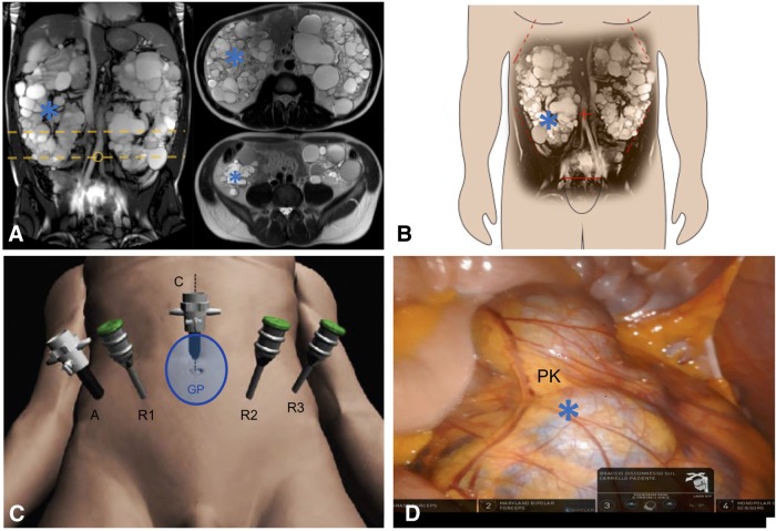
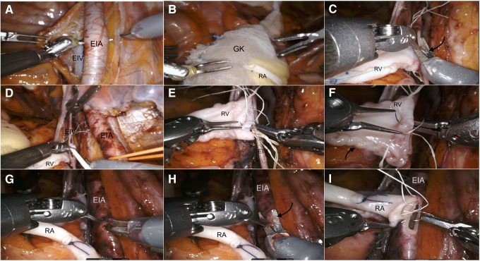
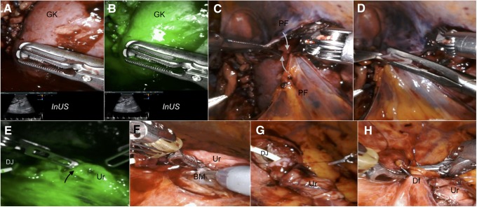
Background: Autosomal dominant polycystic kidney disease (ADPKD) is a common cause of end-stage renal disease (ESRD) and may pose significant technical challenges for kidney transplantation. Recently, robot-assisted kidney transplantation (RAKT) has been shown to achieve excellent patient and graft outcomes while reducing surgical morbidity. However, the vast majority of RAKT performed so far were from living donors and no studies reported the outcomes of RAKT in patients with ADPKD. Case Presentation: Herein, we describe the first successful case of RAKT from a brain-dead deceased donor in a 37-year-old patient with ESRD due to ADPKD. Conclusion: Our case highlights that RAKT can be safely performed by experienced robotic surgeons even in selected complex recipients such as patients with ADPKD and using grafts from deceased donors.

Keywords: autosomal dominant polycystic kidney disease; brain-dead deceased donor; case report; kidney transplantation; robotics.


Conflict of interest statement

The authors declare that they have no conflict of interest.

Figures

<b>FIG. 1.</b>
FIG. 1.
Overview of robotic kidney transplantation in our case. (A, B). MRI images showing the voluminous polycystic native kidneys. No space limitations at the level of transplantation site at the right iliac fossa were noted. The yellow dotted lines in A indicate the level of the transversal sections of the MRI images showed on the right side of A. (C) Trocar placement. A 4- to 5-cm midline periumbilical incision was made for the GelPOINT access device. A pneumoperitoneum of 12 mm Hg was established, and three 8 mm robotic ports and one 12 mm additional assistant port were inserted under vision in a modified RARP configuration. All trocars were positioned ∼3 cm downward on the same lines to obtain an increased working space far from the enlarged PKs. The patient was then turned to a 30° Trendelenburg position and the da Vinci Xi Robot® was docked on the lateral patient side. (D) Intraoperative snapshot showing the distal portion of the enlarged right PK (*): no space constraints were present at the level of the right iliac fossa. PK, polycystic kidney; RARP, robot-assisted radical prostatectomy.
<b>FIG. 2.</b>
FIG. 2.
Intraoperative snapshots showing the step-by-step technique for vascular anastomoses during RAKT in our case. RAKT was performed by a dedicated robotic transplant team. Bench preparation of the graft included defatting, perfusion with Celsior® solution, vessel preparation, and preplacement of a 6F, 14 cm DJ. The graft was then inserted into a gauze jacket filled with ice. (A) Skeletonization of external iliac vessels. The dissection was more extended in our case of RAKT to ensure avoidance of atherosclerotic plaques at the site of arterial anastomosis. (B) Insertion of the graft through the GelPOINT device without need of redocking. (C) Venous anastomosis. A venotomy (arrow) was performed using cold scissors. (D) The lumen of EIV was flushed with heparinized saline before anastomosis. (E, F) A running suture from the 12- to 6-o'clock position was performed to close the posterior plate using the needle driver on the surgeon's dominant hand and the Black diamond microforceps on the nondominant hand (E); then, a knot was tied and the anterior plate was completed with the same suture from the 6- to 12-o'clock position (F). (G, H) Linear arteriotomy and its conversion to circular arteriotomy using cold scissors. (I) Arterial anastomosis. Two half-running sutures starting at the 12 o'clock position and running toward the 6 o'clock position were performed with two threads to close the posterior and then anterior plates of the anastomosis. DJ, Double-J stent; EIA, external iliac artery; EIV, external iliac vein; GK, graft kidney; RA, renal artery; RAKT, robot-assisted kidney transplantation; RV, renal vein.
<b>FIG. 3.</b>
FIG. 3.
Intraoperative snapshots showing graft reperfusion and the key steps of ureterovesical anastomosis in our case. (A, B, E). After venous injection of 0.3 mg/kg of indocyanine green, Firefly® fluorescence imaging technology and InUS were used to check graft reperfusion and integrity of ureteral vascularization (black arrow). (C, D). Closure of the extraperitoneal pouch reapproximating the two previously prepared PF by using hem-o-Lok clips (white arrows). (F–H). Key steps of ureteroneocystostomy using a modified Lich-Gregoir technique. After spatulation, the Ur is anastomosed to the bladder mucosa in a continuous fashion over the preplaced DJ; then, the detrusor muscle was closed with a running suture creating an antirefluxing mechanism. BM, bladder mucosa; Df, detrusor flap; InUS, intraoperative duplex ultrasound; PF, peritoneal flaps; Ur, ureter.

References

    1. Jean RA, Alexandre M, Yoo PS. Kidney transplantation with and without native nephrectomy for polycystic kidney disease: Results of the national inpatient sample and the rationale for a 2-staged procedure. J Am Coll Surg 2018;226:1079–1084 - PubMed
    1. Menon M, Sood A, Bhandari M, et al. . Robotic kidney transplantation with regional hypothermia: A step-by-step description of the Vattikuti Urology Institute–Medanta Technique (IDEAL Phase 2a). Eur Urol 2014;65:991–1000 - PubMed
    1. Marconi L, Figueiredo A. Chapter 14: Cadaver donor organ procurement: Technical aspects and surgical options. In: Figueiredo A, Lledò-Garcìa E, eds. European Texbook on Kidney Tranplantation (ESTU-EAU), 1st ed. 2017. SBN/EAN 9789492671004. © EAU Section Office
    1. Sood A, Ghani KR, Ahlawat R, et al. . Application of the statistical process control method for prospective patient safety monitoring during the learning phase: Robotic kidney transplantation with regional hypothermia (IDEAL phase 2a-b). Eur Urol 2014;66:371–378 - PubMed

Publication types

LinkOut - more resources