. 2018 Dec;23(4):249-267.
doi: 10.1007/s10911-018-9407-1. Epub 2018 Aug 9.

CXCL1 Derived from Mammary Fibroblasts Promotes Progression of Mammary Lesions to Invasive Carcinoma through CXCR2 Dependent Mechanisms

Affiliations

CXCL1 Derived from Mammary Fibroblasts Promotes Progression of Mammary Lesions to Invasive Carcinoma through CXCR2 Dependent Mechanisms

Shira Bernard et al. J Mammary Gland Biol Neoplasia. 2018 Dec.

Abstract

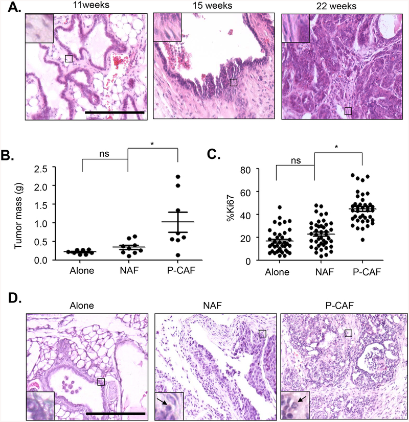
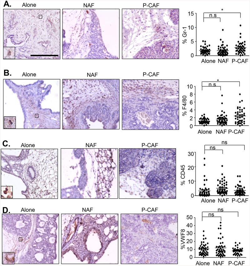
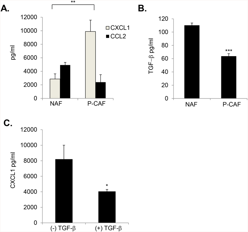
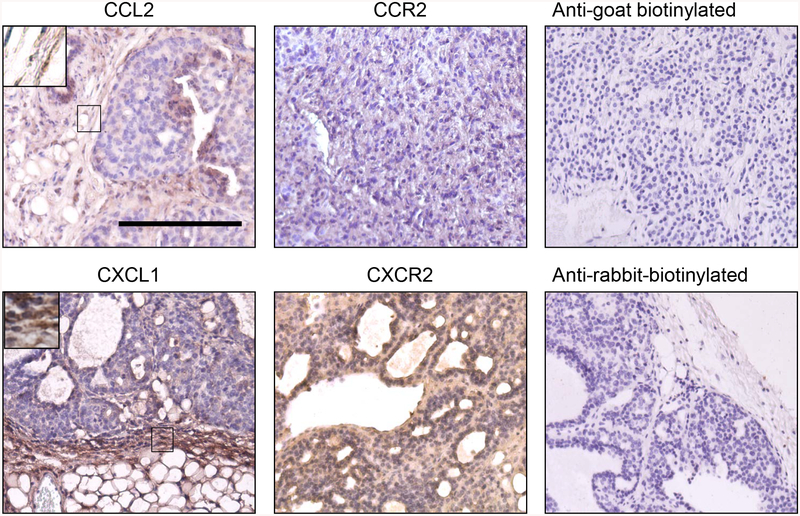
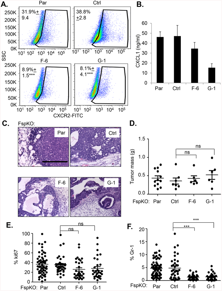
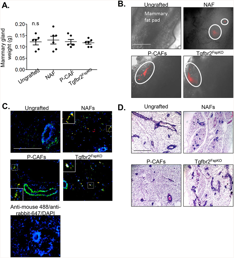
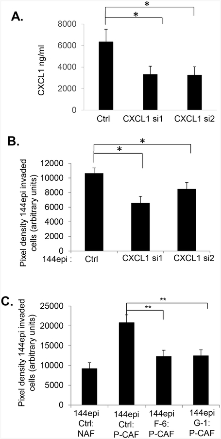
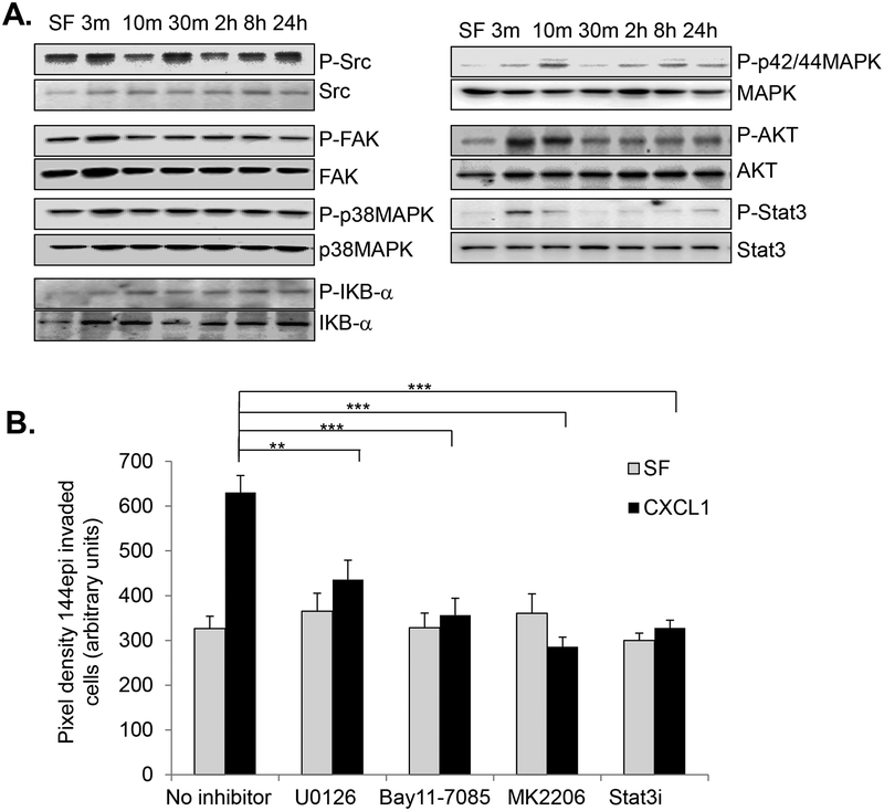
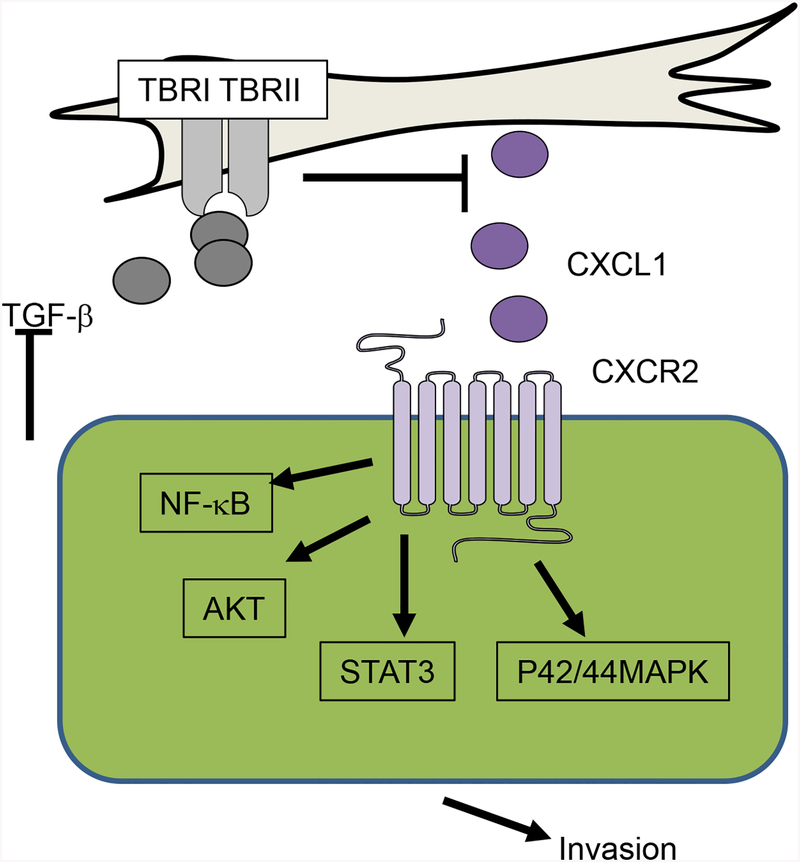
With improved screening methods, the numbers of abnormal breast lesions diagnosed in women have been increasing over time. However, it remains unclear whether these breast lesions will develop into invasive cancers. To more effectively predict the outcome of breast lesions and determine a more appropriate course of treatment, it is important to understand the underlying mechanisms that regulate progression of non-invasive lesions to invasive breast cancers. A hallmark of invasive breast cancers is the accumulation of fibroblasts. Fibroblast proliferation and activation in the mammary gland is in part regulated by the Transforming Growth Factor beta1 pathway (TGF-β). In animal models, TGF-β suppression of CCL2 and CXCL1 chemokine expression is associated with metastatic progression of mammary carcinomas. Here, we show that transgenic overexpression of the Polyoma middle T viral antigen in the mouse mammary gland of C57BL/6 mice results in slow growing non-invasive lesions that progress to invasive carcinomas in a stage dependent manner. Invasive carcinomas are associated with accumulation of fibroblasts that show decreased TGF-β expression and high levels of CXCL1, but not CCL2. Using co-transplant models, we show that decreased TGF-β signaling in fibroblasts contribute to mammary carcinoma progression through enhancement of CXCL1/CXCR2 dependent mechanisms. Using cell culture models, we show that CXCL1 mediated mammary carcinoma cell invasion through NF-κB, AKT, Stat3 and p42/44MAPK dependent mechanisms. These studies provide novel mechanistic insight into the progression of pre-invasive lesions and identify new stromal biomarkers, with important prognostic implications.

Keywords: Breast carcinoma; C57BL/6; Chemokine; Fibroblast; Invasion; MMTV-PyVmT; TGF-β.


Figures

Fig 1.
Fig 1.. P-CAFs enhance progression of 144epi mammary carcinoma cells.
A. H&E stain of mammary lesions from PyVmT/C57BL/6 mice. N=12 for mice 7–11 weeks, n=13 for mice 12–19 weeks old and n=12 for mice 25–29 weeks old. Magnified inset shows level of fibroblastic stroma. B-D. 144epi mammary epithelial cells were transplanted alone, with NAF or P-CAFs for 60 days. Mammary tissues were measured for tumor mass (B), Ki67 immunostaining (C) and H&E stain (D). Magnified inset in (D) shows mammary carcinoma cells. Arrowhead points to invasive carcinoma cells. Statistical analysis was performed using One Way ANOVA with Bonferroni post-hoc comparison. Statistical significance was determined by p<0.05. *p<0.05, **p<0.01. Scale bar=200 microns. n=8 for 144epi alone, n=9 for 144epi: NAF, n=8 for 144epi: P-CAF. Mean±SEM are shown.
Fig 2.
Fig 2.. P-CAFs regulate immune cell recruitment to 144epi mammary lesions.
Mammary lesions were immunostained for: A. Gr-1, B. F4/80, C. CD45 or D. Von Willebrand Factor 8 (VWF8). Statistical analysis was performed using One Way ANOVA with Bonferroni post-hoc comparison. Statistical significance was determined by p<0.05. *p<0.05, n.s=not significant. Scale bar=200 microns. n=8 for 144epi alone, n=9 for 144epi: NAF, n=8 for 144epi: P-CAF. Mean±SEM are shown.
Fig 3.
Fig 3.. P-CAFs show increased CXCL1 and decreased TGF-β expression.
ELISA analysis of A. CXCL1 and CCL2 in NAFs vs. P-CAFs, B. TGF-β in NAFs vs. P-CAFs, C. CXCL1 in P-CAFs treated with 10 ng/ml of TGF-β for 24 hours. Statistical analyzed was performed using Two Tailed T-test. Samples were plated in triplicate per group. Experiments were performed three times. Statistical significance was determined by p<0.05. *p<0.05, **p<0.01, ***p<0.001. Mean±SEM are shown.
Fig 4.
Fig 4.. CXCL1 and CXCR2 are more highly expressed than CCL2 and CCR2 in 144epi mammary lesions.
Mouse mammary lesions from 144epi cells were co-transplanted with P-CAFs and immunostained for CCL2, CCR2, CXCL1 or CXCR2. Magnified insets show staining in fibroblastic cells. Scale bar=200 microns. n=8 per group.
Fig 5.
Fig 5.. CXCR2 knockdown in 144epi cells inhibits Tgfbr2FspKO fibroblasts mediated progression of 144epi mammary lesions.
A. Flow cytometry analysis of CXCR2 in Parental (Par) or 144epi cells stably expressing control shRNA (Ctrl) or CXCR2 shRNAs (F-6, G-1). Percentage of positive cells are shown. B. CXCL1 ELISA of cultured 144epi cells. C-F. Tgfbr2FspKO fibroblasts were co-transplanted with control or CXCR2 deficient 144epi cells. Mammary tissues were analyzed by H&E stain (C), tumor mass (D), Ki67 (E), or Gr-1 expression (F). Statistical analysis was determined by One Way ANOVA followed by Bonferroni post-hoc comparison of F-6 and G-1 with control shRNA. Statistical significance was determined by p <0.05. *p<0.05; **p<0.01, ***p<0.001, ns=not significant. Scale bar=200 microns. n=12 for parental 144epi: Tgfbr2FspKO, n=7 for 144epi: Control shRNA, n=6 for 144epi:F-6, n=6 for 144epi:G-1. Flow cytometry experiments were performed 3 times. Mean±SEM are shown.
Fig 6.
Fig 6.. Fibroblasts transplanted alone do not promote tumor formation in mammary glands.
NAFs, P-CAFs or Tgbr2FspKO fibroblasts were labeled with mcherry and transplanted alone in mammary fat pads of C57Bl/6 mice for up to 60 days. A. Mammary tissues were harvested and imaged by using the FL Auto EVOS imaging system for detection of mCherry positive cells. mCherry positive areas are circled. Scale bar=1000 microns. B. Co-immunofluorescence staining for mCherry (red) or α-sma expression (green) in mammary tissues. Magnified inset shows α-sma/mCherry co-expressing cells. Scale bar=200 microns. C. Mammary tissues were serial sectioned and stained by H&E. Representative of 200 sections per sample, n=6 mice per group. Scale bar=200 microns. Statistical analysis was performed using One Way ANOVA with Bonferroni post-hoc comparison. Statistical significance was determined by p<0.05. ns=not significant. Mean+SEM are shown.
Fig 7.
Fig 7.. Stromal derived CXCL1 promotes 144epi cell invasion through CXCR2 dependent mechanisms.
A-B. P-CAFs were transfected with control (Ctrl) or CXCL1 siRNAs, analyzed for CXCL1 expression by ELISA (A) and 144epi cell invasion by Matrigel transwell assay (B). C. Control (Ctrl) or CXCR2 shRNA expressing144epi cells (G-1, F-6) were co-cultured with control or CXCL1 siRNA transfected P-CAFs and analyzed for changes in invasion by Matrigel transwell assay. Statistical analysis was determined by One Way ANOVA followed by Bonferroni post-hoc analysis. Triplicate samples per group were plated; experiments were performed three times. Statistical significance was determined by p <0.05. *p<0.05; **p<0.01, ns=not significant. Mean±SEM are shown.
Fig 8.
Fig 8.. CXCL1 regulates invasion of 144epi cells through NF-κB, MAPK, AKT and Stat3 dependent pathways.
A. 144epi cells were treated with 60 ng/ml of CXCL1 over time and analyzed by immunoblot for expression of the indicated proteins. B. 144epi cells were treated with 60 ng/ml CXCL1 in the presence or absence of: 5 μM NF-κB inhibitor (Bay11–7082), 1 μM MEK inhibitor (U0126),0.28 μM AKT inhibitor (MK2206) and 1mM Stat3 peptide inhibitors (Stat3i), and analyzed for changes in transwell invasion over 24 hours. Statistical analysis was determined by One Way ANOVA followed by Bonferroni post-hoc analysis. Transwell assays were plated in triplicate per group. Experiments were performed three times. Statistical significance was determined by p <0.05. **p<0.05, ***p<0.001, ns=not significant. Mean±SEM are shown.
Fig 9.
Fig 9.. Role of fibroblasts in invasive progression of breast lesions.
Decreased expression of TGF-β in stromal fibroblasts increase expression of CXCL1, which acts on CXCR2 expressed on mammary epithelial cells. CXCL1/CXCR2 signaling increases activation of: NF-κB, AKT, Stat3 and p42/44MAPK to enhance cellular invasion.

References

    1. Hussain A, Gordon-Dixon A, Almusawy H, Sinha P, Desai A. The incidence and outcome of incidental breast lesions detected by computed tomography. Annals of the Royal College of Surgeons of England. 2010;92(2):124–6. doi:10.1308/003588410X12518836439083. - DOI - PMC - PubMed
    1. Falomo E, Strigel RM, Bruce R, Munoz Del Rio A, Adejumo C, Kelcz F. Incidence and outcomes of incidental breast lesions detected on cross-sectional imaging examinations. Breast J. 2018. doi:10.1111/tbj.13040. - DOI - PubMed
    1. Menes TS, Rosenberg R, Balch S, Jaffer S, Kerlikowske K, Miglioretti DL. Upgrade of high-risk breast lesions detected on mammography in the Breast Cancer Surveillance Consortium. Am J Surg. 2014;207(1):24–31. doi:10.1016/j.amjsurg.2013.05.014. - DOI - PMC - PubMed
    1. Schnitt SJ. Benign breast disease and breast cancer risk: morphology and beyond. The American journal of surgical pathology. 2003;27(6):836–41. - PubMed
    1. Dyrstad SW, Yan Y, Fowler AM, Colditz GA. Breast cancer risk associated with benign breast disease: systematic review and meta-analysis. Breast Cancer Res Treat. 2015;149(3):569–75. doi:10.1007/s10549-014-3254-6. - DOI - PubMed

Publication types

MeSH terms

LinkOut - more resources