Viability of Descemet Membrane Endothelial Keratoplasty Grafts Folded in the Eye Bank
- PMID: 30095494
- PMCID: PMC6173616
- DOI: 10.1097/ICO.0000000000001711
Viability of Descemet Membrane Endothelial Keratoplasty Grafts Folded in the Eye Bank
Abstract
Purpose: Preloaded, trifolded grafts in Descemet membrane endothelial keratoplasty require transfer of the trifolding process from the corneal transplant surgeon to the eye bank technician. We sought to assess whether trifolding may be safely conducted by an eye bank technician with cell loss comparable to standard peeling and lifting.
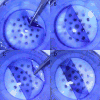
Methods: A total of 10 grafts were stained, peeled, and transferred directly onto a bed of Calcein-AM and Amvisc Plus by an eye bank technician. Five grafts were removed and stained as a scroll, and 5 grafts were trifolded with the endothelium in before transfer. Photographs were acquired with an inverted fluorescence microscope, and image segmentation was performed. A t test was conducted to compare differences in endothelial cell loss across groups.
Results: Mean cell loss in the scroll group was 18.5% [95% confidence interval (CI): 15.2%-21.9%] compared with 7.6% of the trifolded group (95% CI: 1.7%-13.5%). A 2-tailed t test indicated decreased cell loss in the trifolded group (P = 0.013).
Conclusions: Despite additional manipulation of the graft, trifolding of Descemet membrane and endothelium may be performed by an eye bank technician without significantly increased cell loss relative to graft preparation as a scroll.
Conflict of interest statement
Conflict of Interest: The following authors have ownership interest in Treyetech, Inc.: (EC, KB, SC, CC, AS, AE)
Figures


References
-
- Feng MT, Price MO, Price FW., Jr Update on Descemet Membrane Endothelial Keratoplasty (DMEK) Int Ophthalmol Clin. 2013;53:31–45. - PubMed
-
- Dapena I, Ham L, Melles G. Endothelial keratoplasty: DSEK/DSAEK or DMEK - the thinner the better? Curr Opin Ophthalmol. 2009;20:299–307. - PubMed
-
- Tran KD, Dye PK, Odell K, et al. Evaluation and Quality Assessment of Prestripped, Preloaded Descemet Membrane Endothelial Keratoplasty Grafts. Cornea. 2017;36:484–490. - PubMed
-
- Palioura S, Colby K. Outcomes of Descemet Stripping Endothelial Keratoplasty Using Eye Bank-Prepared Preloaded Grafts. Cornea. 2017;36:21–25. - PubMed
MeSH terms
Grants and funding
LinkOut - more resources
Full Text Sources
Other Literature Sources

