

The Recognition of Unrelated Ligands by Identical Proteins
- PMID: 30095890
- PMCID: PMC6499068
- DOI: 10.1021/acschembio.8b00443
The Recognition of Unrelated Ligands by Identical Proteins
Abstract
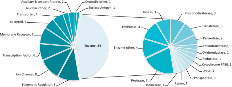
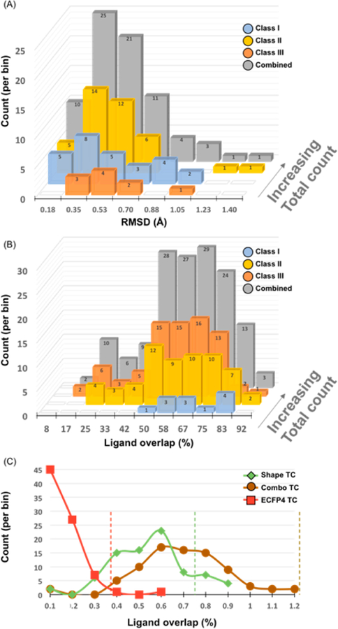
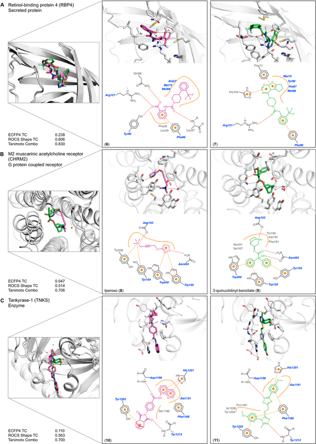
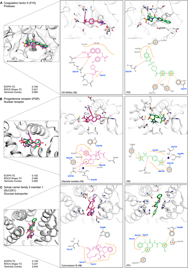
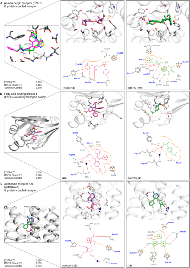
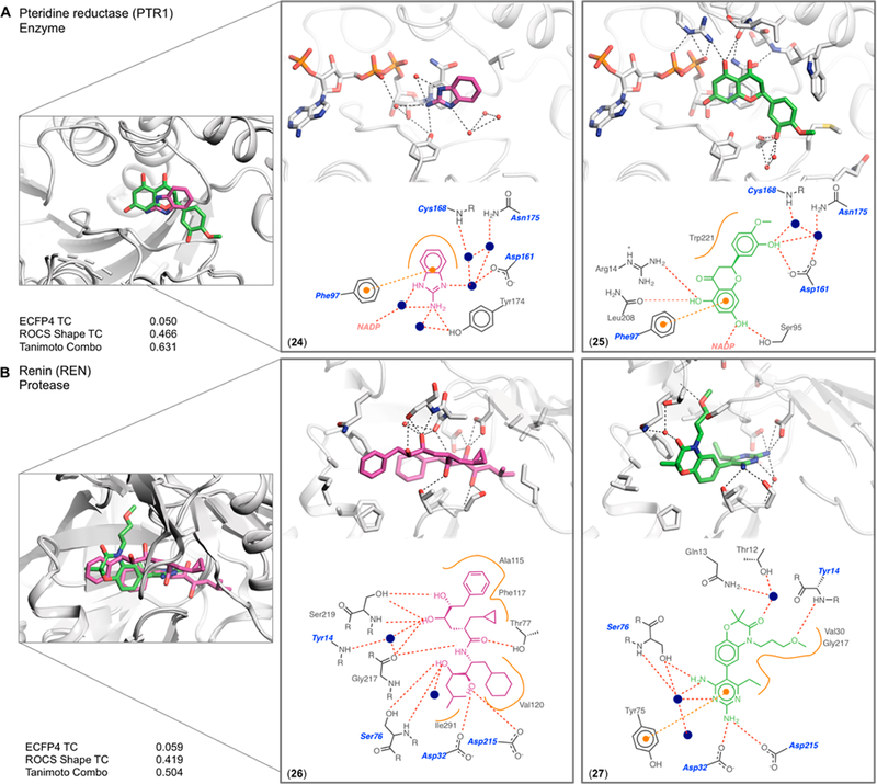
Unrelated ligands, often found in drug discovery campaigns, can bind to the same receptor, even with the same protein residues. To investigate how this might occur, and whether it might be typically possible to find unrelated ligands for the same drug target, we sought examples of topologically unrelated ligands that bound to the same protein in the same site. Seventy-six pairs of ligands, each bound to the same protein (152 complexes total), were considered, classified into three groups. In the first (31 pairs of complexes), unrelated ligands interacted largely with the same pocket residues through different functional groups. In the second group (39 pairs), the unrelated ligand in each pair engaged different residues, though still within the same pocket. The smallest group (6 pairs) contained ligands with different scaffolds but with shared functional groups interacting with the same residues. We found that there are multiple chemically unrelated but physically similar functional groups that can complement any given local protein pocket; when these functional group substitutions are combined within a single molecule, they lead to topologically unrelated ligands that can each well-complement a site. It may be that many active and orthosteric sites can recognize topologically unrelated ligands.
Conflict of interest statement
The authors declare no competing financial interest.
Figures
Similar articles
-
The Recognition of Identical Ligands by Unrelated Proteins.ACS Chem Biol. 2015 Dec 18;10(12):2772-84. doi: 10.1021/acschembio.5b00683. Epub 2015 Oct 12. ACS Chem Biol. 2015. PMID: 26421501 Free PMC article.
-
The use of small-molecule structures to complement protein-ligand crystal structures in drug discovery.Acta Crystallogr D Struct Biol. 2017 Mar 1;73(Pt 3):240-245. doi: 10.1107/S2059798317000675. Epub 2017 Feb 22. Acta Crystallogr D Struct Biol. 2017. PMID: 28291759 Free PMC article.
-
On the importance of composite protein multiple ligand interactions in protein pockets.J Comput Chem. 2017 Jun 5;38(15):1252-1259. doi: 10.1002/jcc.24523. Epub 2016 Nov 16. J Comput Chem. 2017. PMID: 27864975 Free PMC article.
-
New approaches for computing ligand-receptor binding kinetics.Curr Opin Struct Biol. 2018 Apr;49:1-10. doi: 10.1016/j.sbi.2017.10.001. Epub 2017 Nov 11. Curr Opin Struct Biol. 2018. PMID: 29132080 Review.
-
Cryo-EM for Small Molecules Discovery, Design, Understanding, and Application.Cell Chem Biol. 2018 Nov 15;25(11):1318-1325. doi: 10.1016/j.chembiol.2018.07.006. Epub 2018 Aug 9. Cell Chem Biol. 2018. PMID: 30100349 Free PMC article. Review.
Cited by
-
Reversible Covalent Inhibition─Desired Covalent Adduct Formation by Mass Action.ACS Chem Biol. 2024 Apr 19;19(4):824-838. doi: 10.1021/acschembio.3c00805. Epub 2024 Apr 3. ACS Chem Biol. 2024. PMID: 38567529 Free PMC article. Review.
-
Mining the Protein Data Bank to inspire fragment library design.Front Chem. 2023 Feb 10;11:1089714. doi: 10.3389/fchem.2023.1089714. eCollection 2023. Front Chem. 2023. PMID: 36846858 Free PMC article.
-
Pan-HSP90 ligand binding reveals isoform-specific differences in plasticity and water networks.Protein Sci. 2023 May;32(5):e4629. doi: 10.1002/pro.4629. Protein Sci. 2023. PMID: 36938943 Free PMC article.
References
-
- Frimurer TM, Mende F, Graae A-S, Engelstoft MS, Egerod KL, Nygaard R, Gerlach L-O, Hansen JB, Schwartz TW, and Holst B (2017) Model-Based Discovery of Synthetic Agonists for the Zn2+-Sensing G-Protein-Coupled Receptor 39 (GPR39) Reveals Novel Biological Functions. J. Med. Chem 60, 886–898. - PubMed
-
- Huang X-P, Karpiak J, Kroeze WK, Zhu H, Chen X, Moy SS, Saddoris KA, Nikolova VD, Farrell MS, Wang S, Mangano TJ, Deshpande DA, Jiang A, Penn RB, Jin J, Koller BH, Kenakin T, Shoichet BK, and Roth BL (2015) Allosteric ligands for the pharmacologically dark receptors GPR68 and GPR65. Nature 527, 477–483. - PMC - PubMed
-
- Powers RA, Morandi F, and Shoichet BK (2002) Structure-Based Discovery of a Novel, Noncovalent Inhibitor of AmpC β-Lactamase. Structure 10, 1013–1023. - PubMed
-
- Komoriya S, Haginoya N, Kobayashi S, Nagata T, Mochizuki A, Suzuki M, Yoshino T, Horino H, Nagahara T, Suzuki M, Isobe Y, and Furugoori T (2005) Design, synthesis, and biological activity of non-basic compounds as factor Xa inhibitors: SAR study of S1 and aryl binding sites. Bioorg. Med. Chem 13, 3927–3954. - PubMed
Publication types
MeSH terms
Substances
Grants and funding
LinkOut - more resources
Full Text Sources
Other Literature Sources
