. 2018 Aug 8;19(8):2327.
doi: 10.3390/ijms19082327.

Developing Hollow-Channel Gold Nanoflowers as Trimodal Intracellular Nanoprobes

Affiliations

Developing Hollow-Channel Gold Nanoflowers as Trimodal Intracellular Nanoprobes

Sunjie Ye et al. Int J Mol Sci. .

Abstract

Gold nanoparticles-enabled intracellular surface-enhanced Raman spectroscopy (SERS) provides a sensitive and promising technique for single cell analysis. Compared with spherical gold nanoparticles, gold nanoflowers, i.e., flower-shaped gold nanostructures, can produce a stronger SERS signal. Current exploration of gold nanoflowers for intracellular SERS has been considerably limited by the difficulties in preparation, as well as background signal and cytotoxicity arising from the surfactant capping layer. Recently, we have developed a facile and surfactant-free method for fabricating hollow-channel gold nanoflowers (HAuNFs) with great single-particle SERS activity. In this paper, we investigate the cellular uptake and cytotoxicity of our HAuNFs using a RAW 264.7 macrophage cell line, and have observed effective cellular internalization and low cytotoxicity. We have further engineered our HAuNFs into SERS-active tags, and demonstrated the functionality of the obtained tags as trimodal nanoprobes for dark-field and fluorescence microscopy imaging, together with intracellular SERS.

Keywords: SERS; dark-field; fluorescence; gold nanoflowers; intracellular nanoprobes; surface plasmon resonance.


Conflict of interest statement

The authors declare no conflicts of interest.

Figures

Figure 1
Figure 1
(a) UV-vis spectrum of hollow-channel gold nanoflowers (HAuNFs) synthesized with ascorbic acid (AA). (b) TEM image showing multiple branches on the outer surface; (c) TEM image in an orientation showing the hollow channel in the center.
Figure 2
Figure 2
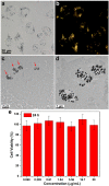
(a) Representative bright-field microscopy and (b) corresponding dark-field microscopy image of RAW macrophage cells treated with HAuNFs. Sectioned TEM images of a fixed Raw 264.7 cell treated with HAuNFs: (c) Low and high (d) magnification. The red arrows in (c) indicate HAuNFs; (e) cell viability of Raw 264.7 cells treated with varying concentrations of HAuNFs. Results are shown as mean ±SD (n = 6) as determined using Cell Counting Kit-8 (CCK-8) assays.
Figure 3
Figure 3
(a) Schematic representation of the surface modification of HAuNFs to form surface-enhanced Raman spectroscopy (SERS) nanotags (HAuNFs@R6G@dBSA); (b) UV-vis spectra of HAuNFs@R6G and HAuNFs@R6G@dBSA; (c) emission spectra (Excitation wavelength: 470 nm) of HAuNFs, HAuNFs@R6G@dBSA, and the aqueous solution of R6G (Inset of c).
Figure 4
Figure 4
Bright-field, dark-field, and fluorescent microscopy images of RAW 264.7 cells after 12 h of incubation in a medium containing HAuNFs@R6G@dBSA, HAuNFs@dBSA, or medium only as a blank control. (Wider-view bright-field microscopy images of cells treated with HAuNFs@R6G@dBSA or HAuNFs@dBSA are shown in Supplementary Materials, Figures S1 and S2.)
Figure 5
Figure 5
Raman spectra of HAuNFs, HAuNFs@dBSA, or HAuNFs@R6G@dBSA on a glass slide and individual Raw 264.7 cells treated with medium containing HAuNFs@dBSA, HAuNFs@R6G@dBSA, or medium only as a blank control. The spectra were recorded under the same measurement conditions, and have been shifted vertically for clarity in comparison. The spectra of nanostructures on a glass slide were collected by drop-casting 20 μL of the sample solution on glass slides and performing the measurements on the concentrated ring area post natural drying.

References

    1. Drescher D., Kneipp J. Nanomaterials in complex biological systems: Insights from Raman spectroscopy. Chem. Soc. Rev. 2012;41:5780–5799. doi: 10.1039/c2cs35127g. - DOI - PubMed
    1. Zong C., Xu M.X., Xu L.J., Wei T., Ma X., Zheng X.S., Hu R., Ren B. Surface-Enhanced Raman Spectroscopy for Bioanalysis: Reliability and Challenges. Chem. Rev. 2018;118:4946–4980. doi: 10.1021/acs.chemrev.7b00668. - DOI - PubMed
    1. Auchinvole C.A.R., Richardson P., McGuinnes C., Mallikarjun V., Donaldson K., McNab H., Campbell C.J. Monitoring Intracellular Redox Potential Changes Using SERS Nanosensors. ACS Nano. 2012;6:888–896. doi: 10.1021/nn204397q. - DOI - PubMed
    1. Vitol E.A., Orynbayeva Z., Friedman G., Gogotsi Y. Nanoprobes for intracellular and single cell surface-enhanced Raman spectroscopy (SERS) J. Raman Spectrosc. 2012;43:817–827. doi: 10.1002/jrs.3100. - DOI
    1. Kneipp K., Haka A.S., Kneipp H., Badizadegan K., Yoshizawa N., Boone C., Shafer-Peltier K.E., Motz J.T., Dasari R.R., Feld M.S. Surface-enhanced Raman Spectroscopy in single living cells using gold nanoparticles. Appl. Spectrosc. 2002;56:150–154. doi: 10.1366/0003702021954557. - DOI