. 2018 Sep;188(9):2016-2024.
doi: 10.1016/j.ajpath.2018.05.010. Epub 2018 Aug 7.

Vasoactive Intestinal Peptide Promotes Corneal Allograft Survival

Affiliations

Vasoactive Intestinal Peptide Promotes Corneal Allograft Survival

Vannarut Satitpitakul et al. Am J Pathol. 2018 Sep.

Abstract

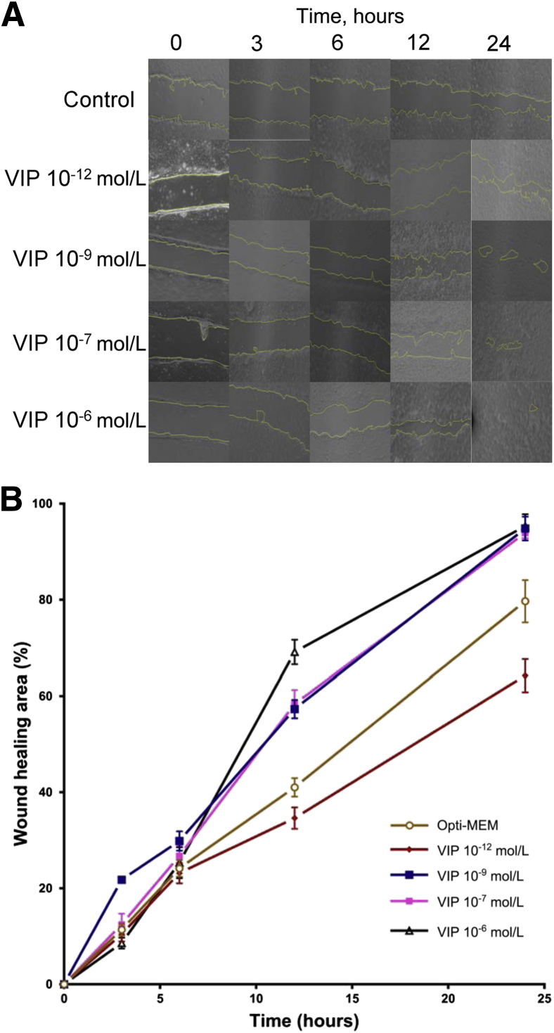
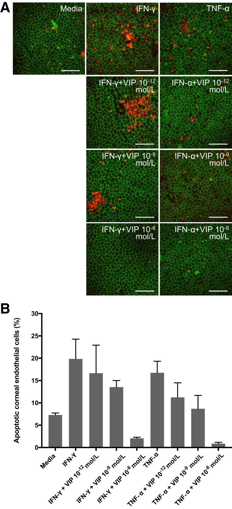
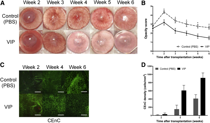
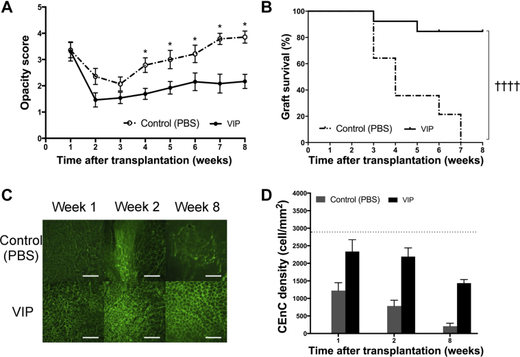
Corneal transplantation is the most prevalent form of tissue transplantation. The success of corneal transplantation mainly relies on the integrity of corneal endothelial cells (CEnCs), which maintain graft transparency. CEnC density decreases significantly after corneal transplantation even in the absence of graft rejection. To date, different strategies have been used to enhance CEnC survival. The neuropeptide vasoactive intestinal peptide (VIP) improves CEnC integrity during donor cornea tissue storage and protects CEnCs against oxidative stress-induced apoptosis. However, little is known about the effect of exogenous administration of VIP on corneal transplant outcomes. We found that VIP significantly accelerates endothelial wound closure and suppresses interferon-γ- and tumor necrosis factor-α-induced CEnC apoptosis in vitro in a dose-dependent manner. In addition, we found that intracameral administration of VIP to mice undergoing syngeneic corneal transplantation with endothelial injury increases CEnC density and decreases graft opacity scores. Finally, using a mouse model of allogeneic corneal transplantation, we found for the first time that treatment with VIP significantly suppresses posttransplantation CEnC loss and improves corneal allograft survival.


Figures

Figure 1
Figure 1
Vasoactive intestinal peptide (VIP) treatment of human corneal endothelial cells accelerates corneal endothelial wound healing in vitro in a dose-dependent manner. A:In vitro wound healing of cultured human corneal endothelial cells with or without VIP (10−12, 10−9, 10−7, 10−6 mol/L). Representative images show the remaining wounded area (yellow) in VIP-treated mice and in the control group (Opti-MEM) 3, 6, 12, and 24 hours after incubation. B: Percentage of healed endothelial area compared with baseline in VIP-treated groups and in controls. Human corneal endothelial cells treated with 10−9, 10−7, and 10−6 mol/L VIP demonstrate significantly enhanced wound healing compared with the control group 12 and 24 hours after incubation compared with the control (P < 0.05, U-test). A dose-dependent trend was observed in the effect of VIP. Data are expressed as means ± SEM and data from one of two independent experiments are shown (B). n = 9. Original magnification, ×40.
Figure 2
Figure 2
Vasoactive intestinal peptide (VIP) suppresses interferon (IFN)-γ– and tumor necrosis factor (TNF)-α–mediated corneal endothelial cell apoptosis. A: Representative confocal micrographs showing naïve C57BL/6 corneal cups incubated with IFN-γ or TNF-α with or without VIP (10−12, 10−9, 10−6 mol/L). After 18 hours of incubation, corneas were stained for zonula occluden-1 (green) and terminal deoxynucleotidyl transferase–mediated dUTP nick-end labeling assay (TUNEL, red) to visualize endothelial cell-to-cell junctions and apoptotic cells, respectively. B: Bar diagram showing the percentages of apoptotic (TUNEL-positive) corneal endothelial cells incubated ex vivo with IFN-γ or TNF-α with different doses of VIP. VIP 10−6 mol/L significantly suppresses IFN-γ– and TNF-α–mediated corneal endothelial cell apoptosis [P = 0.02 and 0.008, respectively (U-test)]. Data are expressed as means ± SEM and data from one of two independent experiments are shown (B). n = 5 corneas (B). Scale bars = 100 μm. Original magnification, ×400.
Figure 3
Figure 3
Vasoactive intestinal peptide (VIP) treatment decreases graft opacity and enhances corneal endothelial wound healing in a murine model of syngeneic corneal transplantation with endothelial injury. A: Representative slit-lamp images showing corneas at week 2 to 6 after transplantation. B: Graft opacity scores are significantly decreased in VIP-treated group from week 2 to 6 after transplantation compared with the control. C: Representative confocal micrographs of central area of transplanted corneas isolated from VIP-treated mice and the control groups at week 2, 4, and 6 after transplantation. To visualize corneal endothelial cell-to-cell junctions, corneas were stained with zonula occluden-1 (green). D: Bar diagram showing central corneal endothelial cell (CEnC) density in VIP-treated corneas compared with control at weeks 2, 4 (P = 0.03), and 6 (P = 0.02) post-after transplantation. Data are expressed as means ± SEM and data from one of two independent experiments are shown. n = 6 corneas (B); n = 15 per group (B). P < 0.05 versus VIP (U-test). Scale bars = 100 μm (C). Original magnification, ×25 (A); ×400 (C). PBS, phosphate-buffered saline.
Figure 4
Figure 4
Effect of vasoactive intestinal peptide (VIP) treatment on high-risk corneal transplant survival. Animals underwent high-risk allogeneic corneal transplantation and received treatment with VIP at 1, 3, 5, 7, and 9 days after transplantation. A: VIP treatment significantly decreases graft opacity scores at 4 to 8 weeks after transplantation. B: Weekly examination of grafts for 8 weeks demonstrates a significant increase in graft survival in VIP-treated mice compared with the controls (85% versus 0%; hazard ratio, 0.10; 95% CI, 0.04–0.26). C: Representative confocal micrographs of central area of transplanted corneas in VIP-treated mice and in controls at 1, 2, and 8 weeks after transplantation. Corneal endothelial cell-to-cell junction were stained and visualized with zonula occluden-1 (green). D: Bar diagram of central corneal endothelial cell (CEnC) densities show significantly higher CEnC density in the VIP-treated group compared with the controls at 8 weeks after transplantation [P = 0.02 (U-test)]. CEnC density in VIP-treated group did not reach statistical significance at weeks 1 and 2 after transplantation compared with the controls (P = 0.11 and P = 0.06, respectively). Horizontal dotted line represents the CEnC density of naïve age-matched C57BL/6 corneas. Data are expressed as means ± SEM and data from one out of two independent experiments are shown. n = 14 (B); n = 5 corneas (D). P < 0.05 versus VIP (U-test); ††††P < 0.0001 (log-rank test). Scale bars = 100 μm (C). Original magnification, ×400 (C). PBS, phosphate-buffered saline.

References

    1. Gain P., Jullienne R., He Z., Aldossary M., Acquart S., Cognasse F., Thuret G. Global survey of corneal transplantation and eye banking. JAMA Ophthalmology. 2016;134:167–173. - PubMed
    1. Niederkorn J.Y. The immune privilege of corneal allografts. Transplantation. 1999;67:1503–1508. - PubMed
    1. The Collaborative Corneal Transplantation Studies Research Group The collaborative corneal transplantation studies (CCTS): effectiveness of histocompatibility matching in high-risk corneal transplantation. Arch Ophthalmol. 1992;110:1392–1403. - PubMed
    1. Obata H., Ishida K., Murao M., Miyata K., Sawa M. Corneal endothelial cell damage in penetrating keratoplasty. Jpn J Ophthalmol. 1991;35:411–416. - PubMed
    1. Acar B.T., Vural E.T., Acar S. Changes in endothelial cell density following penetrating keratoplasty and deep anterior lamellar keratoplasty. Int Journal Ophthalmol. 2011;4:644–647. - PMC - PubMed

Publication types

Substances