. 2018 Oct 1;217(10):3656-3669.
doi: 10.1083/jcb.201804028. Epub 2018 Aug 10.

Reconstitution reveals Ykt6 as the autophagosomal SNARE in autophagosome-vacuole fusion

Affiliations

Reconstitution reveals Ykt6 as the autophagosomal SNARE in autophagosome-vacuole fusion

Levent Bas et al. J Cell Biol. .

Abstract

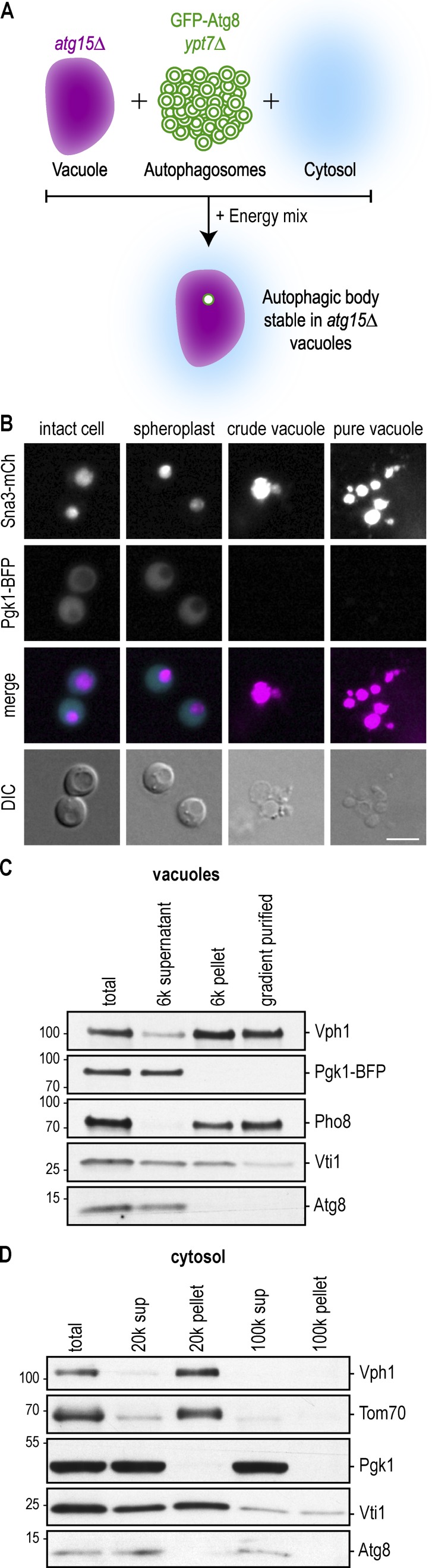
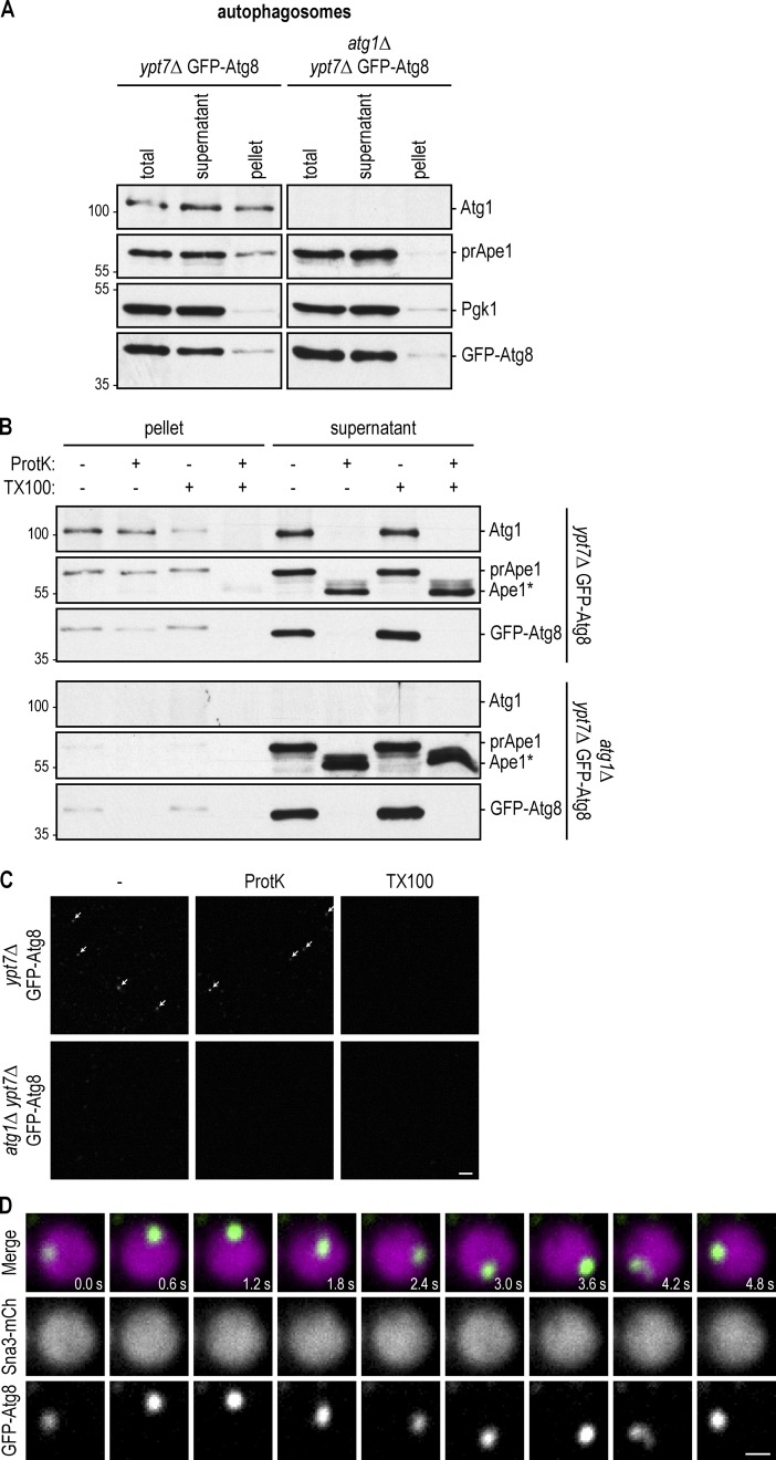
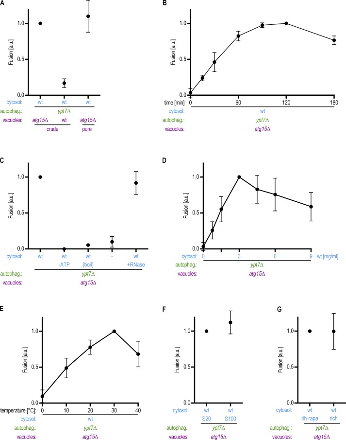
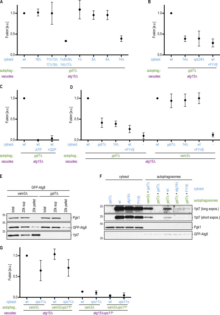
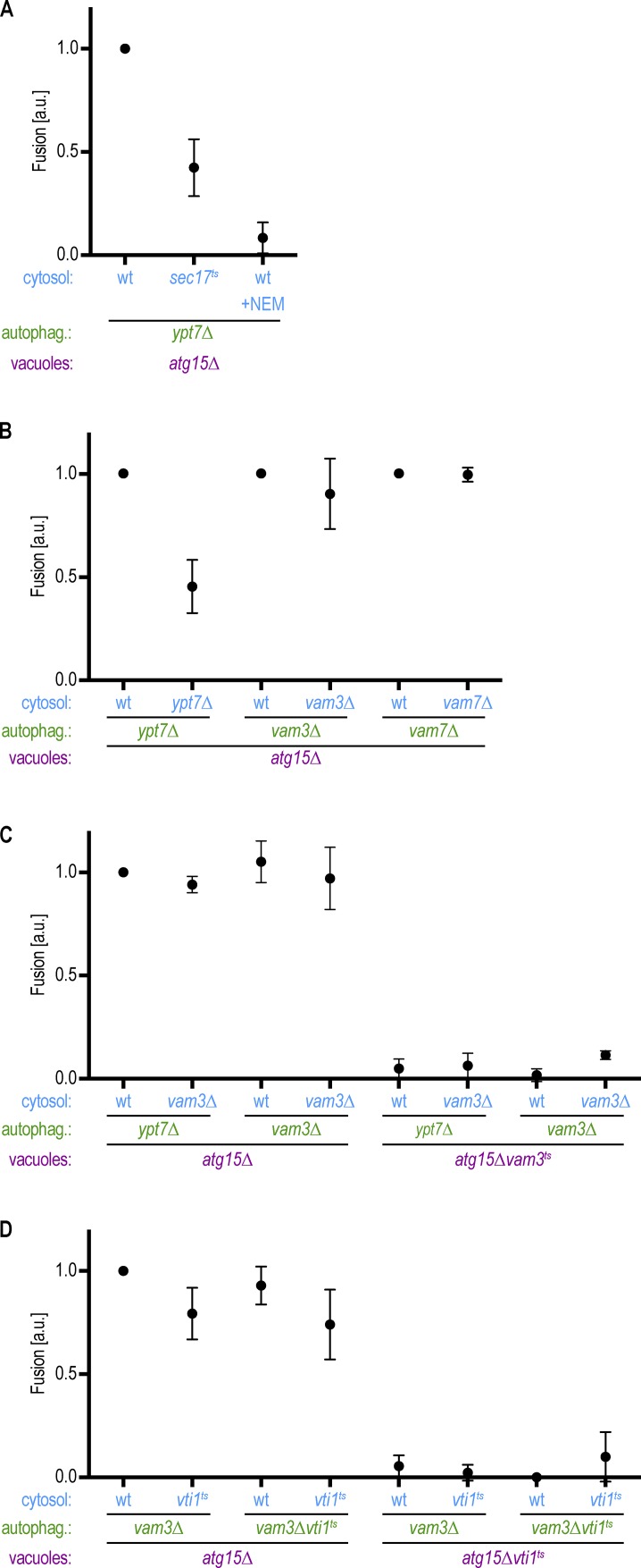
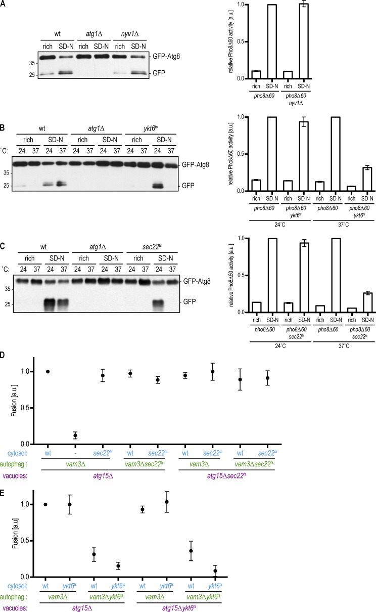
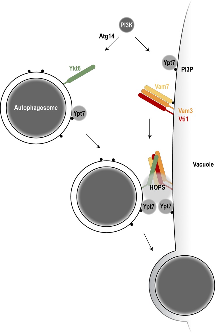
Autophagy mediates the bulk degradation of cytoplasmic material, particularly during starvation. Upon the induction of autophagy, autophagosomes form a sealed membrane around cargo, fuse with a lytic compartment, and release the cargo for degradation. The mechanism of autophagosome-vacuole fusion is poorly understood, although factors that mediate other cellular fusion events have been implicated. In this study, we developed an in vitro reconstitution assay that enables systematic discovery and dissection of the players involved in autophagosome-vacuole fusion. We found that this process requires the Atg14-Vps34 complex to generate PI3P and thus recruit the Ypt7 module to autophagosomes. The HOPS-tethering complex, recruited by Ypt7, is required to prepare SNARE proteins for fusion. Furthermore, we discovered that fusion requires the R-SNARE Ykt6 on the autophagosome, together with the Q-SNAREs Vam3, Vam7, and Vti1 on the vacuole. These findings shed new light on the mechanism of autophagosome-vacuole fusion and reveal that the R-SNARE Ykt6 is required for this process.


Figures

Figure 1.
Figure 1.
Preparation of cytosolic and vacuolar fractions. (A) Schematic of the experimental setup: crude vacuolar, autophagosomal, and cytosolic fractions are individually prepared from yeast cells. Incubation of these three fractions together with an energy regeneration system allows the fusion of autophagosomes with vacuoles in vitro. (B) Vacuoles were isolated from Sna3-mCherry Pgk1-BFP atg15Δ cells. Logarithmically growing cells were harvested and lysed, and vacuoles were separated from the cytosolic fraction by a 6,000 g spin. The pellet containing the vacuoles was then further separated on a 0–4–8–15% Ficoll step gradient, and vacuoles were collected at the 0–4% Ficoll interface. The purification steps were analyzed by fluorescence microscopy. DIC, differential interference contrast. Bar, 5 µm. (C) The individual fractions from B were analyzed by anti-Vph1, anti-Pgk1, anti-Pho8, anti-Vti1, and anti-Atg8 Western blotting. One representative experiment out of three is shown. (D) Cytosol was prepared from WT cells treated with 220 nM rapamycin for 4 h by freezer milling and centrifugation at 20,000 g. The supernatant was further spun at 100,000 g. Fractions were analyzed by anti-Vph1, anti-Tom70, anti-Pgk1, anti-Vti1, and anti-Atg8 Western blotting. One representative experiment out of three is shown. Molecular masses are given in kilodaltons.
Figure 2.
Figure 2.
Reconstitution of autophagosome–vacuole fusion in vitro. (A) GFP-Atg8 ypt7Δ cells were starved for 16 h. After cell lysis, autophagosomes were enriched in a 20,000 g pellet and analyzed by anti-Atg1, anti-Ape1, anti-Pgk1, and anti-GFP Western blotting. atg1Δypt7Δ cells that cannot form autophagosomes served as a control. One representative experiment out of three is shown. (B) Samples from A were subjected to proteinase K (ProtK) and Triton X-100 (TX100) treatment as indicated and analyzed by anti-Atg1, anti-Ape1, and anti-GFP Western blotting. Ape1*, proteinase K–resistant fragment of Ape1. One representative experiment out of three is shown. Molecular masses are given in kilodaltons. (C) Samples from A were subjected to proteinase K and Triton X-100 treatment as indicated and analyzed by fluorescence microscopy. Arrows point to autophagosomes. One representative experiment out of three is shown. Bar, 10 µm. (D) The autophagosomal, vacuolar, and cytosolic fractions from Figs. 1 (C and D) and 2 A were coincubated together with an energy regeneration system for 2 h. Fusion was monitored by fluorescence microscopy and judged by the appearance of a mobile green dot in the vacuole. Shown are stills of a 12-s time-lapse video (Video 1). Bar, 0.5 µm.
Figure 3.
Figure 3.
Autophagosome–vacuole fusion depends on time, temperature, and cytosol concentration and requires energy. (A) Vacuoles containing or lacking Atg15 as indicated were analyzed in an in vitro fusion assay as described in Fig. 2 D. (B) Samples as described in Fig. 2 D were incubated for the time indicated, and autophagosome–vacuole fusion was quantified. (C) Fusion reactions were performed in the presence and absence of an ATP regeneration system and with or without the prior incubation of the cytosolic fraction for 5 min at 95°C (boil) or RNase treatment. Dash indicates that no cytosol was added. (D) The amount of cytosol added to the fusion reaction was titrated as indicated. (E) Fusion reactions were performed at different temperatures as indicated. (F) Comparison of a 20,000 g and a 100,000 g cytosolic supernatant in the fusion reaction. (G) Comparison of cytosol prepared from logarithmically growing cells (rich) with cytosol from cells that had been treated with 220 nm rapamycin for 4 h before harvesting (rapa). All graphs show the mean from at least three independent experiments. Error bars are SD.
Figure 4.
Figure 4.
The PI3-kinase complex I is required for the recruitment of Ypt7 to autophagosomes to promote autophagosome–vacuole fusion. (A) Fusion assay as in Fig. 2 D. Cytosol was prepared from the indicated deletion strains. (B) Fusion assay as in Fig. 2 D. Cytosol was prepared from atg14Δ or vps34Δ cells or from cells overexpressing the FYVE domain. (C) Fusion assay as in Fig. 2 D. Fusion reactions were either mock treated or incubated without an ATP regeneration system or with 10 mM GDP. (D) Fusion assay as in Fig. 2 D. Cytosol and autophagosomes were prepared from the indicated deletion strains. The FYVE domain was overexpressed where indicated. (E) Autophagosomes were isolated from the indicated strains as in Fig. 2 A. The presence of Ypt7 in the autophagosome-enriched pellet was monitored by Western blotting. One representative experiment out of three is shown. (F) Autophagosomes were isolated from the indicated strains as in Fig. 2 A, incubated with cytosolic fractions as indicated, and again isolated by centrifugation. The binding of Ypt7 to the autophagosomes was assessed by Western blotting. One representative experiment out of three is shown. Molecular masses are given in kilodaltons. (G) Fusion assay as in Fig. 2 D. Cytosol, autophagosomes, and vacuoles were prepared from the indicated deletion or ts strains at 24°C. Fusion reactions were incubated at the restrictive temperature (30°C) for 2 h. All graphs show the mean from at least three independent experiments. Error bars are SD.
Figure 5.
Figure 5.
The Q-SNAREs Vam3, Vam7, and Vti1 act on the vacuole to promote autophagosome–vacuole fusion. (A) Fusion assay as in Fig. 2 D. Cytosol was prepared from the indicated deletions strains. The Sec18 inhibitor 5 mM N-ethylmaleimide (NEM) was added where indicated. (B) Fusion assay as in Fig. 2 D. Cytosol and autophagosomes were prepared from the indicated deletions strains. (C and D) Fusion assays as in Fig. 2 D. Cytosol, autophagosomes, and vacuoles were prepared from the indicated deletion or ts strains at 24°C. Fusion reactions were incubated at the restrictive temperature (30°C) for 2 h. All graphs show the mean from three independent experiments. Error bars are SD.
Figure 6.
Figure 6.
The R-SNARE Ykt6 acts on the autophagosome to promote autophagosome–vacuole fusion. (A) Left: The indicated strains expressing GFP-Atg8 were grown to logarithmic phase (rich) and were subsequently starved for 4 h (SD-N). TCA extracts were prepared, and GFP-Atg8 cleavage was monitored by anti-GFP Western blotting. One representative experiment out of three is shown. Right: Pho8Δ60 assay of nyv1Δ cells. Indicated cells were grown to mid–log phase and starved for 4 h where indicated. Pho8Δ60-specific alkaline phosphatase activity was measured in three independent experiments, and the mean was plotted normalized to starved pho8Δ60 alkaline phosphatase activity. (B and C) Left: The indicated strains expressing GFP-Atg8 were grown at 24°C to logarithmic phase. Cultures were split and incubated for 4 h at the permissive (24°C) or restrictive (37°C) temperature, with (SD-N) or without starvation (rich) as indicated. TCA extracts were prepared, and GFP-Atg8 cleavage was monitored by anti-GFP Western blotting. One representative experiment out of three is shown. Right: Pho8Δ60 assay. The indicated strains were grown at 24°C to logarithmic phase. Cultures were split and incubated for 4 h at the permissive (24°C) or restrictive (37°C) temperature, with (SD-N) or without starvation (rich) as indicated. Pho8Δ60-specific alkaline phosphatase activity was measured in three independent experiments, and the mean was plotted normalized to starved pho8Δ60 alkaline phosphatase activity at the respective temperature. Molecular masses are given in kilodaltons. (D and E) Fusion assay as in Fig. 2 D. Cytosol, autophagosomes, and vacuoles were prepared from the indicated deletion or ts strains at 24°C. Fusion reactions were incubated at the restrictive temperature (30°C) for 2 h. All graphs show the mean from at least three independent experiments. Error bars are SD.
Figure 7.
Figure 7.
Model of autophagosome–vacuole fusion. Atg14 in the PI3-kinase complex I is required to produce PI3P on the autophagosome to allow Ypt7 recruitment. The R-SNARE Ykt6 localizes to autophagosomes; the Q-SNAREs Vam3, Vam7, and Vti1 localize to the vacuole. With the help of Ypt7 on both the autophagosome and the vacuole and the HOPS tethering complex, trans-SNARE complex formation is promoted and allows autophagosome–vacuole fusion.

References

    1. Abeliovich H., Darsow T., and Emr S.D.. 1999. Cytoplasm to vacuole trafficking of aminopeptidase I requires a t-SNARE-Sec1p complex composed of Tlg2p and Vps45p. EMBO J. 18:6005–6016. 10.1093/emboj/18.21.6005 - DOI - PMC - PubMed
    1. Araki Y., Ku W.C., Akioka M., May A.I., Hayashi Y., Arisaka F., Ishihama Y., and Ohsumi Y.. 2013. Atg38 is required for autophagy-specific phosphatidylinositol 3-kinase complex integrity. J. Cell Biol. 203:299–313. 10.1083/jcb.201304123 - DOI - PMC - PubMed
    1. Cabrera M., and Ungermann C.. 2008. Purification and in vitro analysis of yeast vacuoles. Methods Enzymol. 451:177–196. 10.1016/S0076-6879(08)03213-8 - DOI - PubMed
    1. Cheever M.L., Sato T.K., de Beer T., Kutateladze T.G., Emr S.D., and Overduin M.. 2001. Phox domain interaction with PtdIns(3)P targets the Vam7 t-SNARE to vacuole membranes. Nat. Cell Biol. 3:613–618. 10.1038/35083000 - DOI - PubMed
    1. Chen Y., Zhou F., Zou S., Yu S., Li S., Li D., Song J., Li H., He Z., Hu B., et al. . 2014. A Vps21 endocytic module regulates autophagy. Mol. Biol. Cell. 25:3166–3177. 10.1091/mbc.e14-04-0917 - DOI - PMC - PubMed

Publication types

MeSH terms

Substances

LinkOut - more resources