. 2019 Jan;27(1):206-214.
doi: 10.1007/s00167-018-5090-4. Epub 2018 Aug 10.

Predicting meniscal tear stability across knee-joint flexion using finite-element analysis

Affiliations

Predicting meniscal tear stability across knee-joint flexion using finite-element analysis

Angela E Kedgley et al. Knee Surg Sports Traumatol Arthrosc. 2019 Jan.

Abstract

Purpose: To analyse the stress distribution through longitudinal and radial meniscal tears in three tear locations in weight-bearing conditions and use it to ascertain the impact of tear location and type on the potential for healing of meniscal tears.

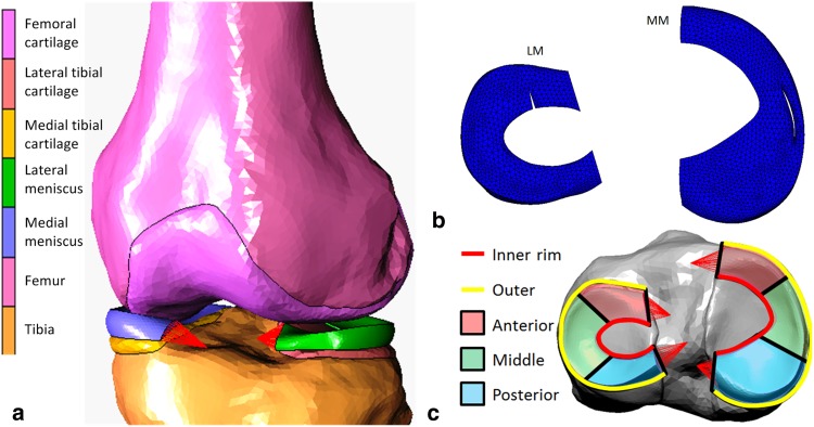
Methods: Subject-specific finite-element models of a healthy knee under static loading at 0°, 20°, and 30° knee flexion were developed from unloaded magnetic resonance images and weight-bearing, contrast-enhanced computed tomography images. Simulations were then run after introducing tears into the anterior, posterior, and midsections of the menisci.

Results: Absolute differences between the displacements of anterior and posterior segments modelled in the intact state and those quantified from in vivo weight-bearing images were less than 0.5 mm. There were tear-location-dependent differences between hoop stress distributions along the inner and outer surfaces of longitudinal tears; the longitudinal tear surfaces were compressed together to the greatest degree in the lateral meniscus and were most consistently in compression on the midsections of both menisci. Radial tears resulted in an increase in stress at the tear apex and in a consistent small compression of the tear surfaces throughout the flexion range when in the posterior segment of the lateral meniscus.

Conclusions: Both the type of meniscal tear and its location within the meniscus influenced the stresses on the tear surfaces under weight bearing. Results agree with clinical observations and suggest reasons for the inverse correlation between longitudinal tear length and healing, the inferior healing ability of medial compared with lateral menisci, and the superior healing ability of radial tears in the posterior segment of the lateral meniscus compared with other radial tears. This study has shown that meniscal tear location in addition to type likely plays a crucial role in dictating the success of non-operative treatment of the menisci. This may be used in decision making regarding conservative or surgical management.

Keywords: Arthroscopy; Biomechanics; Finite element analysis; Knee; Meniscal tear; Meniscus; Reconstruction; Repair; Surgery.


Conflict of interest statement

Conflict of interest

The authors declare that they have no conflict of interest.

Ethical approval

This article does not contain any studies with human participants or animals performed by any of the authors.

Figures

Fig. 1
Fig. 1
a Finite-element model of the tibiofemoral joint. The red lines represent the meniscal insertional ligaments. b Example of an unstable radial tear at the anterior region of the lateral meniscus and an unstable full-thickness vertical longitudinal tear at the middle region of the medial meniscus. c Anterior, middle, and posterior regions of the menisci
Fig. 2
Fig. 2
a Contour map of minimum principal value of stress (MPa) for tibial cartilage at 0°, 20°, and 30° flexion. b Tensor plots of maximum principal value of stress (MPa) in the menisci at 0°, 20°, and 30° flexion. The purple outlines show the unloaded positions of the menisci. The arrows show the direction of displacement from the unloaded positions. The red dashed outlines show approximately the region where hoop stress is dominant
Fig. 3
Fig. 3
Meniscal hoop stress, measured as maximum principal value of stress, is sampled from the inner (normalised length = 0) to the outer (normalised length = 1) rim at the anterior, middle and posterior regions of the lateral and medial menisci
Fig. 4
Fig. 4
Representative (a, c) tensor plots and (b, d) maximum principal value of stress (max PCS) sampled from the inner (normalised length = 0) to the outer (normalised length = 1) rim for longitudinal (a, b) stable and (c, d) unstable tears in the posterior segment of the medial meniscus at 30° of knee flexion. Dashed arrows represent hoop stress. Solid arrows represent the component of the stress tensor acting radially inwards. Thicker arrows represent higher magnitudes
Fig. 5
Fig. 5
Differences in hoop stress, represented by the maximum principal stress, between the surfaces of a 7 mm longitudinal tear in all regions of both menisci plotted against flexion angle
Fig. 6
Fig. 6
Representative a tensor plot for an unstable radial tear and b maximum principal value of stress (max PCS) sampled from the inner (normalised length = 0) to the outer (normalised length = 1) rim for stable and unstable tears in the posterior segment of the medial meniscus for 0° knee flexion. Differences between the high stresses at the tear apex and the intact condition are indicated by the red brackets
Fig. 7
Fig. 7
Difference in maximum principal stress at the apex of a stable radial tear in all regions of both menisci plotted against flexion angle. Positive values indicate tension and negative values indicate compression

References

    1. Baratz ME, Fu FH, Mengato R. Meniscal tears: the effect of meniscectomy and of repair on intraarticular contact areas and stress in the human knee. A preliminary report. Am J Sports Med. 1986;14:270–275. doi: 10.1177/036354658601400405. - DOI - PubMed
    1. Barrett GR, Field MH, Treacy SH, Ruff CG. Clinical results of meniscus repair in patients 40 years and older. Arthroscopy. 1998;14:824–829. doi: 10.1016/S0749-8063(98)70018-0. - DOI - PubMed
    1. Bell JS, Winlove CP, Smith CW, Dehghani H. Modeling the steady-state deformation of the solid phase of articular cartilage. Biomaterials. 2009;30:6394–6401. doi: 10.1016/j.biomaterials.2009.08.026. - DOI - PubMed
    1. Belzer J, Cannon W. Meniscus tears: treatment in the stable and unstable knee. J Am Acad Orthop Surg. 1993;1:41–47. doi: 10.5435/00124635-199309000-00006. - DOI - PubMed
    1. Boxheimer L, Lutz AM, Treiber K, Goepfert K, Crook DW, Marincek B, Weishaupt D. MR imaging of the knee: position related changes of the menisci in asymptomatic volunteers. Invest Radiol. 2004;39:254–263. doi: 10.1097/01.rli.0000116895.04239.84. - DOI - PubMed

Publication types