. 2018 Oct;110(2):239-261.
doi: 10.1111/mmi.14100. Epub 2018 Oct 3.

Rhodobacterales use a unique L-threonine kinase for the assembly of the nucleotide loop of coenzyme B12

Affiliations

Rhodobacterales use a unique L-threonine kinase for the assembly of the nucleotide loop of coenzyme B12

Norbert K Tavares et al. Mol Microbiol. 2018 Oct.

Abstract

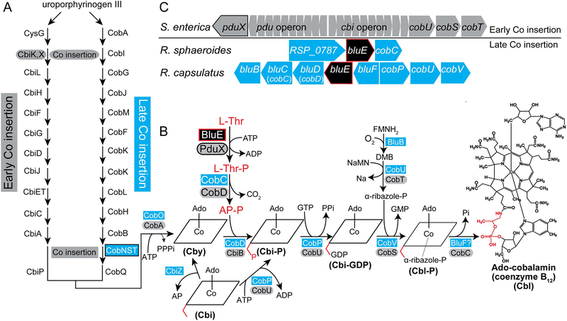
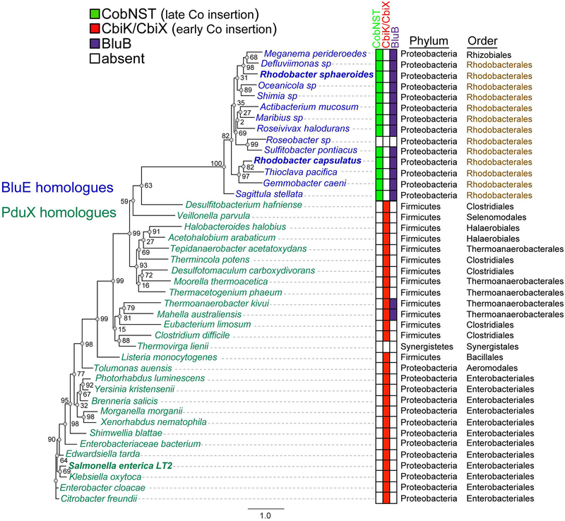
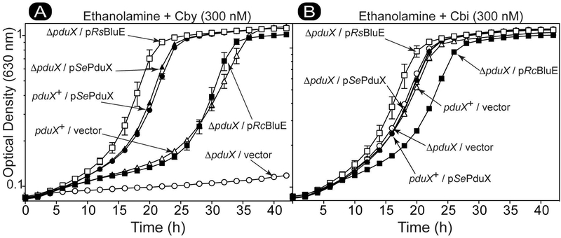
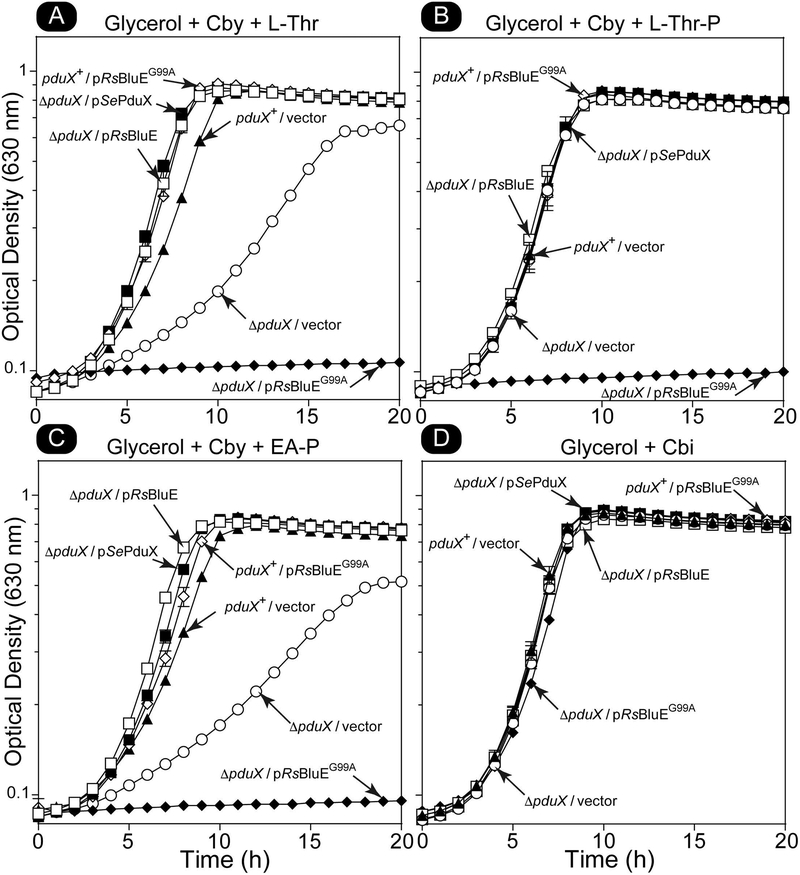
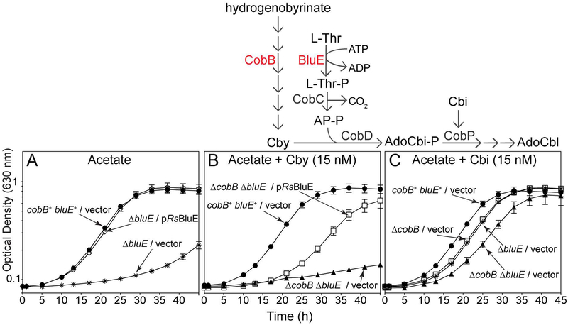
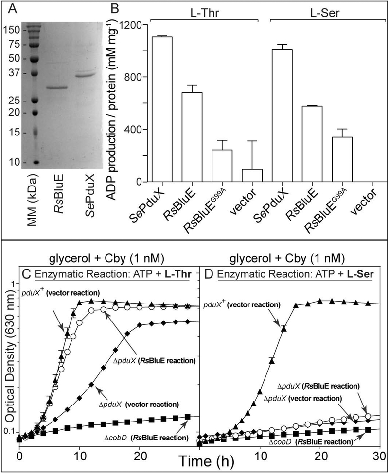
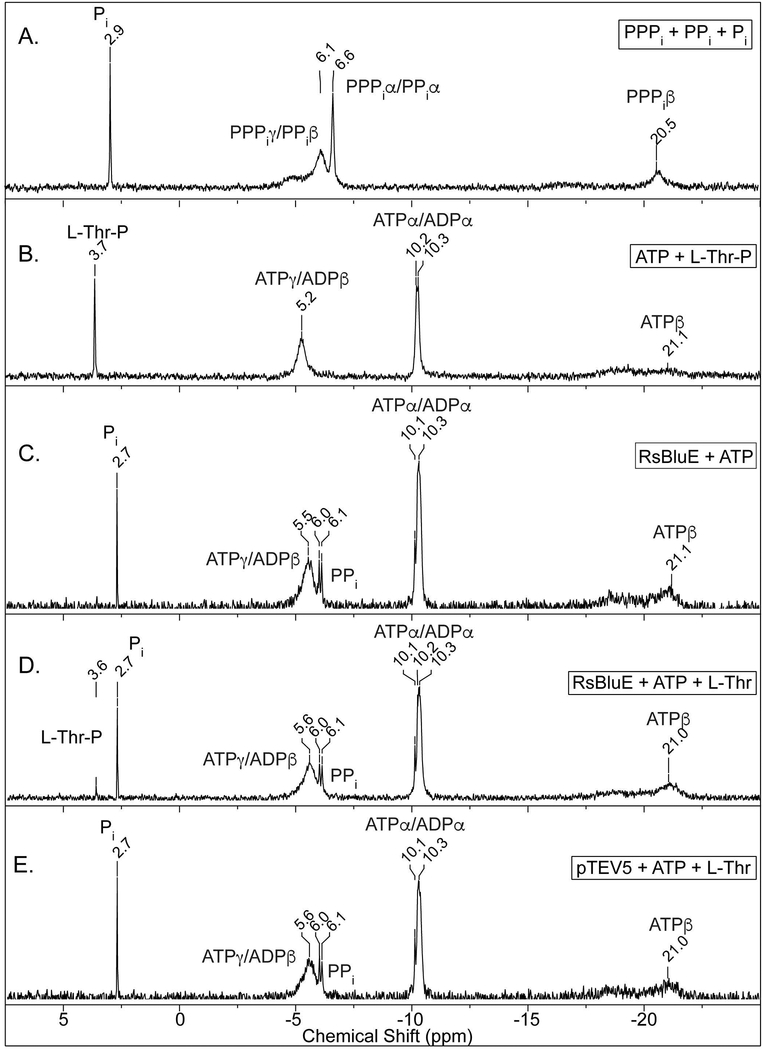
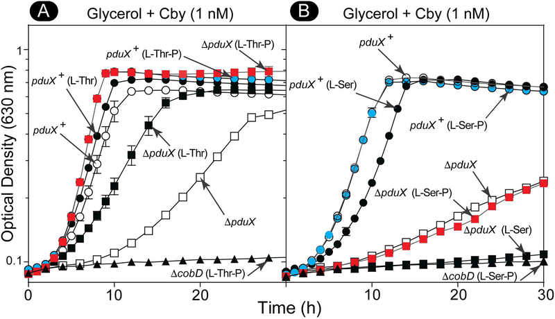
Several of the enzymes involved in the conversion of adenosylcobyric acid (AdoCby) to adenosylcobamide (AdoCba) are yet to be identified and characterized in some cobamide (Cba)-producing prokaryotes. Using a bioinformatics approach, we identified the bluE gene (locus tag RSP_0788) of Rhodobacter sphaeroides 2.4.1 as a putative functional homolog of the L-threonine kinase enzyme (PduX, EC 2.7.1.177) of S. enterica. In AdoCba, (R)-1-aminopropan-2-ol O-phosphate (AP-P) links the nucleotide loop to the corrin ring; most known AdoCba producers derive AP-P from L-Thr-O-3-phosphate (L-Thr-P). Here, we show that RsBluE has L-Thr-independent ATPase activity in vivo and in vitro. We used 31 P-NMR spectroscopy to show that RsBluE generates L-Thr-P at the expense of ATP and is unable to use L-Ser as a substrate. BluE from R. sphaeroides or Rhodobacter capsulatus restored AdoCba biosynthesis in S. enterica ΕpduX and R. sphaeroides ΕbluE mutant strains. R. sphaeroides ΕbluE strains exhibited a decreased pigment phenotype that was restored by complementation with BluE. Finally, phylogenetic analyses revealed that bluE was restricted to the genomes of a few Rhodobacterales that appear to have a preference for a specific form of Cba, namely Coᴽ-(ᴽ-5,6-dimethylbenzimidazolyl-Coᵦ-adenosylcobamide (a.k.a. adenosylcobalamin, AdoCbl; coenzyme B12 , CoB12 ).


Figures

Figure 1.
Figure 1.. AdoCbl biosynthetic pathways in bacteria and archaea.
A. Schematic depicting the early- and late-cobalt insertion pathways of AdoCbl biosynthesis. B. Late steps of the AdoCbl biosynthetic pathway, with proteins in the early-cobalt-insertion (a.k.a. O2-independent, blue rectangles) and late-cobalt-insertion (a.k.a. O2-dependent, gray ovals) pathways. The BluE enzyme is shown in a black box with red trim. The (R)-1-aminopropan-2-ol O-phosphate (AP-P) moiety is highlighted in red in the structure scheme. Exogenous Cbi (a.k.a (CN)2Cbi) enters the pathway as indicated. Exogenous Cby (a.k.a (CN)2Cby) enters the pathway at the location it appears on in the figure. Exogenous corrinoids, including complete Cbas such as Cbl are adenylated by CobA/CobO upon transport into the cell and prior to entering the pathway. C. Genetic layout of the L-threonine kinase-encoding genes bluE in the late-cobalt-insertion, AdoCbl-synthesizing bacteria R. sphaeroides and R. capsulatus, and pduX in S. enterica, an early-cobalt-insertion, AdoCbl-synthesizing bacterium. Figure key: Ado, Adenosyl; Cby, cobyric acid; Cbi-P, cobinamide phosphate; Cbi-GDP, cobinamide-GDP; Cbl-P, cobalamin phosphate; Cbl, cobalamin; AP-P, (R)-1-aminopropan-2-ol O-phosphate; AP, (R)-1-aminopropan-2-ol; L-Thr-P, L-threonine-O-3-phosphate; L-Thr, L-threonine; α-ribazole-P, α-ribazole phosphate; DMB, 5,6-dimethylbenzimidazole; NaMN, nicotinic acid mononucleotide; Nm, Nicotinic Acid; PPi, pyrophosphate; Pi, orthophosphate; CbiK/CbiX, anaerobic cobaltochelatase; CobN/CobS/CobT, aerobic hydrogenobyrinic acid a,c-diamide cobaltochelatase; CobA/CobO, ATP:Co(I)rrinoid Adenosyltransferase; CbiB/CobD, AdoCbi-P synthase; CobS/CobV, AdoCba-5′P synthase; CobD/CobC, L-Thr-P decarboxylase; CobT/CobU, NaMN:DMB phosphoribosyltransferase; CobU/CobP, AdoCbi kinase / AdoCbi-P guanylyltransferase; PduX/BluE, L-Thr kinase; CobC/BluF, AdoCba-5′-P phosphatase; CbiZ, cobinamide amidohydrolase; pdu, genes of the Cba-dependent 1,2-propanediol degradation operon; cbi, genes of the early steps of the early-Co-insertion cobalamin biosynthesis operon.
Figure 2.
Figure 2.. Sequence alignment of representative PduX and BluE proteins.
Genera names of organisms with BluE homologues are colored blue and PduX homologues are colored black. Residues that are 100% conserved are highlighted in red with white lettering. Areas with a high degree of conservation greater than 75% but less than 100% and or residues with similar properties are boxed in blue with red letter.
Figure 3.
Figure 3.. Phylogenetic analysis of the distribution of PduX and BluE proteins.
Maximum likelihood phylogenetic tree of homologous proteins based on the amino acid sequence of SePduX (dark green) and RsBluE or RcBluE (blue). Order Rhodobacterales is highlighted in brown. Color-coded table of the presence or absence of the cobaltochelatase CbiK or CbiX (red squares), which is indicative of the early-cobalt-insertion pathway, the presence of all three subunits of the cobaltochelatase CobNST (lime green squares) in organisms that use the late-cobalt-insertion pathway and BluB (purple squares) the O2-dependent DMB synthase, which is indicative of a physiological reliance on Cbas with DMB as the lower ligand base. Sequences were aligned using the MUSCLE (Edgar, 2004) plugin within Geneious R8.1.7 software (Biomatters Ltd.) with 100 iterations and default settings. Maximum likelihood phylogenic tree was generated with the online PhyML (Guindon et al., 2010) tool on the ATCG Montpellier Bioinformatics Platform available at http://www.atgc-montpellier.fr/phyml/, using Jones-Taylor-Thornton substitution model (Jones et al., 1992) with 500 bootstrap replicates. Bootstrap support for each node is shown as a percent value. The scale bar is provided as a reference for branch lengths. Gene locus tags are available in Table S1.
Figure 4.
Figure 4.. RsBluE and RcBluE restore AdoCbl synthesis in a S. enterica ΔpduX strain.
Growth analysis of S. enterica pduX+ and ΔpduX strains harboring plasmids expressing bluE+ from R. sphaeroides (R.s. bluE+), R. capsulatus (R.c. bluE+), PduX from S. enterica (S.e. pduX+), or containing the empty vector pBAD24 (vector). Cells grown aerobically at 37°C in NCE minimal medium with ethanolamine (90 mM) as the sole carbon and energy source, supplemented with DMB (0.15 mM), arabinose (0.5 mM), ampicillin (0.1 mg mL−1), MgSO4 (1 mM), Fe(III)-citrate (0.05 mM), and A. Cby (300 nM) or B. Cbi (300 nM). A representative graph of three independent growth experiments performed in technical triplicate. Error bar represent the standard error of the mean. Figure key: ΔpduX/pRsBluE (□), ΔpduX/pSePduX (▲), ΔpduX/pRcBluE (■), ΔpduX/vector (○), pduX+/pSePduX (●), pduX+/vector (△).
Figure 5.
Figure 5.. RsBluEG99A variant disrupts all the enzymes in the entire AP-P synthesis and attachment branch in S. enterica in vivo.
Representative graphs of growth analyses of S. enterica cells grown aerobically at 37°C in NCE minimal medium with glycerol (22 mM) ampicillin (0.1 mg mL−1), and MgSO4 (1 mM) Cby (1 nM) and supplemented with A. L-Thr (1 mM), B. L-Thr-P (1 mM), or C. ethanolamine phosphate (EA-P, 1 mM). Cbi (1 mM) was used as the corrinoid in place of Cby in panel D. Experiments were replicated in two independent experiments, each performed in triplicate. Error bar represent the standard error of the mean. Figure key: ΔpduX/pRsBluE (□), ΔpduX/pSePduX (■), ΔpduX/pRsBluEG99A (◆), pduX+/pRsBluEG99A (◇), pduX+/vector (▲), ΔpduX/vector (○).
Figure 6.
Figure 6.. Growth analysis of R. sphaeroides ΔbluE and ΔbluE ΔcobB strains.
Growth analysis of R. sphaeroides bluE+, ΔbluE, and ΔcobB ΔbluE strains harboring a plasmid expressing R. sphaeroides bluE+ or carrying the empty pBBR1MCS-2 plasmid (vector). Cells were grown overnight in Sistrom’s medium and cultures were prepared as described in Materials and Methods. Cell growth was monitored normoxically at 30°C in Sistrom’s medium with acetate (30 mM), kanamycin (0.01 mg mL−1), and when noted, Cby (15 nM) or Cbi (15 nM). Growth experiments were performed in triplicate in three independent experiments. Error bars represent the standard error of the mean. Pathway represents a simplified schematic of the roles of CobB (hydrogenobyrinate a,c-diamide synthase) and BluE (L-Thr kinase) in the synthesis of cobalamin in R. sphaeroides. CobD, AdoCbi-P synthase; CobC, L-Thr-P decarboxylase; CobP, AdoCbi kinase / AdoCbi-P guanylyltransferase; BluE, L-Thr kinase; Cby, cobyric acid; Cbi, cobinamide, AP-P, (R)-1-aminopropan-2-ol O-phosphate. Figure key: Panel A: cobB+ bluE+/vector (●), ΔbluE/pRsBluE (○), ΔbluE/vector (✲), Panel B: ΔcobB ΔbluE/pRsBluE (□), ΔcobB ΔbluE/vector (▲), cobB+ bluE+/pRsBluE (●), Panel C: cobB+ bluE+/vector (●), ΔcobB ΔbluE/vector (▲), ΔcobB/vector (□), ΔbluE/vector (✲).
Figure 7.
Figure 7.. In vitro ATPase activity assay for RsBluE.
A. SDS-PAGE-gel of purified, sarkosyl-solubilized RsBluE and SePduX. B. ATPase activity assay performed with ADP-Glo™ ATPase Assay kit (Promega). Reaction mixtures contained HEPES buffer (50 mM, pH 7.0 at 25°C), MgCl2 (1 mM), ATP (0.1 mM), L-Thr or L-Ser (10 mM), enrichment samples of SePduX, RsBluE, and RsBluEG99A sarkosyl-solubilized protein (12 μM) incubated at 25°C for 1 h. Negative control reaction mixtures contained extracts of sarkosyl-solubilized protein obtained from cells expressing the empty overexpression vector pTEV5 (vector). ATP to ADP conversion was quantified from the luminescence (relative light units; RLU) after subtracting the background from the no-enzyme control and comparing the value to a standard curve (Fig. S3). Graph titles indicate the growth medium and the substrate constituents of the reaction mixtures used to supplement the growth medium. The source of the protein extracts used in the reactions are in parentheses in bold typeface next to the strain genotype (RsBluE reaction or vector resection). Representative graphs of two independent experiments performed in triplicate. Error bars represent the standard deviation from the mean. C and D. Growth analysis of S. enterica cells grown normoxically at 37°C in NCE minimal medium with glycerol (22 mM), MgSO4 (1 mM), Cby (1 nM), and filter sterilized RsBluE reaction mixtures (6% v/v) described above (reactions from panel B) containing either ATP (0.08 mM) and L-Thr (0.8 mM, panel C) or L-Ser (0.8 mM, panel D). The final concentrations of substrates after dilution of the filtered reaction mixtures into the medium are in parentheses. Graphs are representative of two independent experiments performed in triplicate. Error bars represent the standard error of the mean. Figure key: Panel C, D: (▲) pduX+ strain supplemented with filter sterilized negative control enzymatic reaction containing ATP, and L-Thr (panel C) or L-Ser (panel D) and sarkosyl-solubilized protein extracts from cells carrying the empty vector; (◆) ΔpduX strain supplemented with filter sterilized negative control enzymatic reaction containing ATP, and L-Thr (panel C) or L-Ser (panel D) and sarkosyl-solubilized protein extracts from cells carrying the empty vector; (○) ΔpduX strain supplemented with filtered enzymatic reaction containing ATP, and L-Thr (panel C) or L-Ser (panel D) and purified sarkosyl-solubilized RsBluE protein; (■) ΔcobD strain supplemented with filtered enzymatic reaction containing ATP and L-Thr (panel C) or L-Ser (panel D) and purified sarkosyl-solubilized RsBluE protein.
Figure 8.
Figure 8.. 31P-NMR spectra of RsBluE reaction products.
Representative 31P-NMR spectra of duplicate independent experiments. Reaction mixtures containing MgCl2 (1 mM), ATP (3 mM), L-Thr (6 mM), and 10 μL of detergent-free RsBluE protein (11 μM) were incubated at 25°C for 1 h. A. No enzyme reactions with AMP, sodium ortho-phosphate (Pi), sodium pyrophosphate (PPi), and sodium polytriphosphate (PPPi) standards. B. No enzyme reactions with L-Thr-P and ATP standards. C. Reaction containing ATP and RsBluE. D. Reaction containing ATP, L-Thr, and RsBluE. E. Reaction containing ATP, L-Thr, and extracts from cells carrying pTEV5 empty vector. Conditions used for the acquisition of the spectra are described in the Materials and Methods section.
Figure 9.
Figure 9.. Growth analysis of S. enterica strains in the presence of L-Thr, L-Thr-P, L-Ser, and L-Ser-P.
Growth analysis of S. enterica cells grown normoxically at 37°C in NCE minimal medium with glycerol (22 mM), MgSO4 (1 mM), and A. Cby (5 nM) supplemented with L-Thr or L-Thr-P (1 mM), or B. Cby (1 nM) supplemented with L-Ser or L-Ser-P (1 mM). The amino acid or phospho-amino acid supplement is indicated in parentheses next to the strain genotype. Representative graphs of two independent experiments performed in triplicate. Error bars represent the standard error of the mean. Figure key: Panel A: ΔpduX (□), ΔpduX (L-Thr) (■), ΔpduX (L-Thr-P) (■), pduX+ (○), pduX+ (L-Thr) (●), pduX+ (L-Thr-P) (●), ΔcobD (L-Thr-P) (▲), Panel B: ΔpduX (□), ΔpduX (L-Ser) (■), ΔpduX (L-Ser-P) (■), pduX+ (○), pduX+ (L-Ser) (●), pduX+ (L-Ser-P) (●), ΔcobD (L-Ser-P) (▲).

Similar articles

Cited by

References

    1. Altschul SF, Madden TL, Schaffer AA, Zhang J, Miller W, and Lipmann DJ (1997) Gapped BLAST and PSI-BLAST: a new generation of protein database search programs. Nucl. Acids Res 25: 3389–3402. - PMC - PubMed
    1. Amin SA, Green DH, Gardes A, Romano A, Trimble L, and Carrano CJ (2012) Siderophore-mediated iron uptake in two clades of Marinobacter spp. associated with phytoplankton: the role of light. Biometals 25: 181–192. - PubMed
    1. Anderson PJ, Lango J, Carkeet C, Britten A, Kräutler B, Hammock BD, and Roth JR (2008) One pathway can incorporate either adenine or dimethylbenzimidazole as an alpha-axial ligand of B12 cofactors in Salmonella enterica. J. Bacteriol 190: 1160–1171. - PMC - PubMed
    1. Andersson DI, and Roth JR (1989) Mutations affecting regulation of cobinamide biosynthesis in Salmonella typhimurium. J. Bacteriol 171: 6726–6733. - PMC - PubMed
    1. Balch WE, and Wolfe RS (1976) New approach to the cultivation of methanogenic bacteria: 2-mercaptoethanesulfonic acid (HS-CoM)-dependent growth of Methanobacterium ruminantium in a pressurized atmosphere. Appl. Environ. Microbiol 32: 781–791. - PMC - PubMed

Publication types

MeSH terms

LinkOut - more resources