. 2019 Apr:77:41-57.
doi: 10.1016/j.matbio.2018.08.004. Epub 2018 Aug 8.

Prostate cancer sheds the αvβ3 integrin in vivo through exosomes

Affiliations

Prostate cancer sheds the αvβ3 integrin in vivo through exosomes

Shiv Ram Krishn et al. Matrix Biol. 2019 Apr.

Abstract

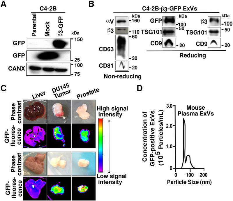
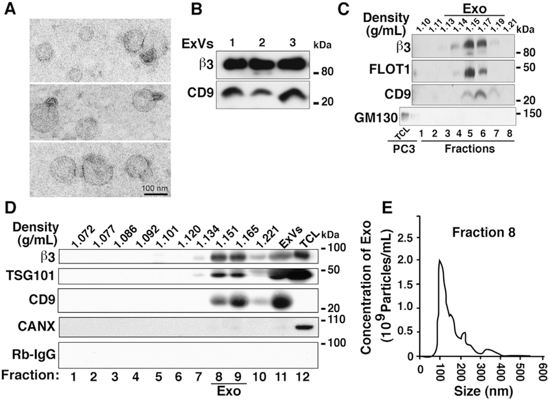
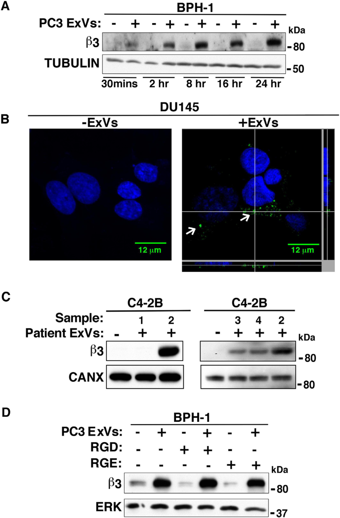
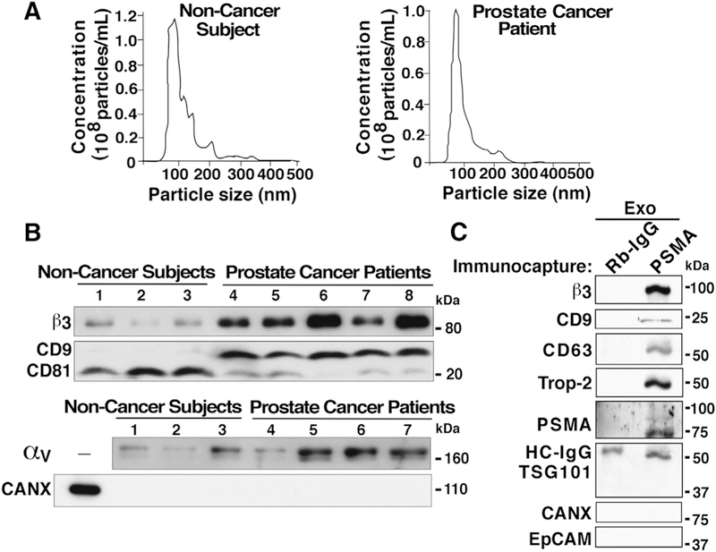
The αvβ3 integrin has been shown to promote aggressive phenotypes in many types of cancers, including prostate cancer. We show that GFP-labeled αvβ3 derived from cancer cells circulates in the blood and is detected in distant lesions in NOD scid gamma (NSG) mice. We, therefore, hypothesized that αvβ3 travels through exosomes and tested its levels in pools of vesicles, which we designate extracellular vesicles highly enriched in exosomes (ExVs), and in exosomes isolated from the plasma of prostate cancer patients. Here, we show that the αvβ3 integrin is found in patient blood exosomes purified by sucrose or iodixanol density gradients. In addition, we provide evidence that the αvβ3 integrin is transferred through ExVs isolated from prostate cancer patient plasma to β3-negative recipient cells. We also demonstrate the intracellular localization of β3-GFP transferred via cancer cell-derived ExVs. We show that the ExVs present in plasma from prostate cancer patients contain higher levels of αvβ3 and CD9 as compared to plasma ExVs from age-matched subjects who are not affected by cancer. Furthermore, using PSMA antibody-bead mediated immunocapture, we show that the αvβ3 integrin is expressed in a subset of exosomes characterized by PSMA, CD9, CD63, and an epithelial-specific marker, Trop-2. Finally, we present evidence that the levels of αvβ3, CD63, and CD9 remain unaltered in ExVs isolated from the blood of prostate cancer patients treated with enzalutamide. Our results suggest that detecting exosomal αvβ3 integrin in prostate cancer patients could be a clinically useful and non-invasive biomarker to follow prostate cancer progression. Moreover, the ability of αvβ3 integrin to be transferred from ExVs to recipient cells provides a strong rationale for further investigating the role of αvβ3 integrin in the pathogenesis of prostate cancer and as a potential therapeutic target.

Keywords: Abiraterone acetate; Enzalutamide; Extracellular vesicles; Plasma exosomes; Prostate cancer; αvβ3 integrin.


Figures

Fig. 1.
Fig. 1.
GFP-tagged αvβ3 integrin is expressed in prostate cancer cell ExVs, and it is localized in distant lesions in vivo. (A) IB analysis of expression of GFP in total cell lysates (TCL) from parental C4-2B, C4-2B-GFP (Mock) and C4-2B-β3-GFP cells. Calnexin (CANX) was used as loading control. (B) IB analysis of expression of αv, β3, CD63, CD81 (non-reducing conditions) and GFP, β3, TSG101, CD9 (reducing conditions) in lysates from ExVs derived from C4-2B-β3-GFP cells by differential ultracentrifugation. (C) In vivo fluorescence and corresponding phase contrast images are shown for liver, DU145 tumor, and prostate isolated from NSG mice that were injected subcutaneously with C4-2B-β3-GFP cells on the right side and non-transfected DU145 cells on the left side. Two representative samples are shown. (D) NTA for size distribution and concentration of GFP-positive ExVs isolated by differential ultracentrifugation from plasma of the mice subcutaneously injected with C4-2B-β3-GFP cells. One representative sample is shown.
Fig. 2.
Fig. 2.
Expression of αvβ3 integrin in exosomes derived from plasma of prostate cancer patients. (A) Transmission electron microscopy (TEM) of negatively stained ExVs isolated from prostate cancer patient plasma by differential ultracentrifugation. Scale bar = 100 nm. (B) IB analysis for expression of p3 integrin and exosomal marker CD9 in lysates from ExVs purified from prostate cancer patient plasma by differential ultracentrifugation. The results from three representative samples are shown. (C) Sucrose gradient analysis of ExVs isolated from prostate cancer patient serum using Exoquick™. Expression of β3 integrin and exosomal markers FLOT1 and CD9 in eight different fractions is shown. GM130 (cis-Golgi marker) is expressed in PC3 lysate (TCL) and is absent in all fractions. The density at which exosomes float in sucrose gradient is between 1.13 and 1.19 g/mL. (D) IB analysis for expression of β3 integrin in ten fractions derived from iodixanol gradient centrifugation of ExVs isolated by differential ultracentrifugation of prostate cancer patient plasma. Expression of TSG101 and CD9 was analyzed as markers present in exosomes, Calnexin (CANX) was analyzed as a marker absent in exosomes, while Rabbit IgG (Rb-IgG) was used as a negative control for β3. ExV lysate derived by ultracentrifugation was used as input and PC3 lysate (TCL) was used as positive control for expression of β3, TSG101, and CANX. (E) NTA for size distribution and concentration of purified exosomes (Exo) in fraction eight (Density = 1.151 g/mL) from iodixanol density gradient.
Fig. 3.
Fig. 3.
ExVs mediate transfer of αvβ3 integrin to recipient cells. (A) IB analysis shows β3 expression levels in BPH-1 cells incubated with PC3-derived ExVs (+) at different time points (30 min to 24 h). BPH-1 cells treated with PBS alone (–) are used as a negative control. TUBULIN expression is included as loading control. (B) DU145 cells were incubated with ExVs (40 μg/ml, right) derived from C4–2B-β3-GFP cells or vehicle alone (left) for 24 h. An intracellular green fluorescent signal corresponding to ExV mediated internalization of β3-GFP was evaluated by confocal microscopy. DAPI was used to detect cell nuclei (blue). Scale bar = 12 μm. The GFP signal corresponding to ExV mediated internalization of β3-GFP in DU145 cells is shown (white arrows). Z-stack analysis shows the intracellular GFP signal in a cell. (C) ExVs were isolated from the plasma of 4 patients (designated 1, 2, 3 and 4) by ultracentrifugation and incubated with C4–2B cells. After 24 h, IB was used to analyze β3 integrin expression levels. C4–2B cells treated with vehicle alone are used as a negative control. CANX serves as loading control. (D) PC3-derived ExVs pre-treated with GRGDSPK peptide (1 mg/mL) for 1 h at 4 °C were incubated with serum-starved BPH-1 cells for 24 h followed by IB analysis to measure β3 levels. BPH-1 cells incubated with GRGESP (1 mg/mL) are used as a negative control. ERK is used as loading control.
Fig. 4.
Fig. 4.
Size distribution analysis and differences in β3 integrin levels in ExVs from plasma of prostate cancer patients compared to subjects not affected by cancer. (A) NTA of ExVs isolated by differential ultracentrifugation from plasma of individuals not affected by cancer (left panel) and prostate cancer patients (right panel). (B) IB analysis of β3, CD9, CD81, and αv levels in ExVs isolated by differential ultracentrifugation from plasma of prostate cancer patients compared to age-matched individuals not affected by cancer. CANX was analyzed as a marker absent in exosomes. Lanes 1, 2, and 3: EV lysates isolated after the plasma was pooled from at least two subjects not affected by cancer (total of 7 biological samples represented in 3 lanes); lanes 4–8: EV lysates from individual patients. 30 μg of exosome lysates were loaded in each lane. (C) lodixanol gradient purified Exosomes (Exo) from prostate cancer patient plasma (pooled from n = 3) were immunocaptured with an antibody to Prostate Specific Membrane Antigen (PSMA) or isotype rabbit immunoglobulin (Rb-IgG) conjugated with Dynabeads M-270 epoxy magnetic beads, according to the manufacturer’s protocol. The immunocaptured whole exosomes were lysed with RIPA buffer, and lysates were separated by SDS-PAGE (7.5% gel). IB analysis shows expression of β3, CD9 and CD63 (exosomal markers), Trop-2, and PSMA; in contrast, TSG101 (exosomal marker), CANX and EpCAM were not detected. HC-IgG, heavy chain IgG.
Fig. 5.
Fig. 5.
Gene expression analysis of CD9 and CD81 in prostate cancer compared to normal samples in publicly available gene expression profiling datasets. (A–D): Gene expression boxplots for CD9. (A) TCGA dataset-prostate adenocarcinoma versus normal. (B) Wallace dataset- prostate adenocarcinoma vs. normal. (C) Lapointe dataset- prostate carcinoma vs. normal. (D) Yu dataset-prostate carcinoma vs. normal. (E-H): Gene expression boxplots for CD81. (E) TCGA dataset-prostate adenocarcinoma vs. normal. (F) Vanaja dataset-prostate adenocarcinoma vs. normal. (G) Taylor dataset-prostate carcinoma vs. normal. (H) Singh dataset- prostate carcinoma vs. normal. 1, Normal; 2, Carcinoma or Adenocarcinoma.
Fig. 6.
Fig. 6.
Nanoparticle tracking analysis (NTA) of ExVs and PSA levels in serum from prostate cancer patients. (A) The size distribution analysis and concentrations were determined for patients in the following groups: patients that were not treated with abiraterone acetate or enzalutamide (Non-treated), treated with abiraterone acetate only (A only), and treated with enzalutamide only (E only). (B) PSA levels in serum of prostate cancer patients, before and after androgen deprivation therapy (abiraterone acetate or enzalutamide).
Fig. 7.
Fig. 7.
β3 integrin levels in plasma of patients treated with enzalutamide (E) compared to patients non-treated (NT) with this drug. IB analysis reveals expression levels of β3 integrin in ExVs derived by differential ultracentrifugation of plasma from patients either non-treated (NT) or treated with enzalutamide (E). CD9, CD63 serve as markers enriched in exosomes and CANX as a marker absent in exosomes. TCL from PC3 cells was used as a positive control for the expression of β3, CD63, CD9, and CANX.

References

    1. Siegel RL, Miller KD, Jemal A, Cancer statistics, 2018, CA Cancer J. Clin. 68 (1) (2018) 7–30. - PubMed
    1. Mitchell T, Neal DE, The genomic evolution of human prostate cancer, Br. J. Cancer 113 (2) (2015) 193–198. - PMC - PubMed
    1. Culig Z, Targeting the androgen receptor in prostate cancer, Expert. Opin. Pharmacother. 15 (10) (2014) 1427–1437. - PubMed
    1. Isaacs JT, The biology of hormone refractory prostate cancer. Why does it develop? Urol. Clin. North Am. 26 (2) (1999) 263–273. - PubMed
    1. Beer TM, Armstrong AJ, Rathkopf DE, Loriot Y, Sternberg CN, Higano CS, Iversen P, Bhattacharya S, Carles J, Chowdhury S, Davis ID, de Bono JS, Evans CP, Fizazi K, Joshua AM, Kim CS, Kimura G, Mainwaring P, Mansbach H, Miller K, Noonberg SB, Perabo F, Phung D, Saad F, Scher HI, Taplin ME, Venner PM, Tombal B, Enzalutamide in metastatic prostate cancer before chemotherapy, N. Engl. J. Med. 371 (5) (2014) 424–433. - PMC - PubMed

Publication types

MeSH terms