. 2018 Aug 11;15(1):224.
doi: 10.1186/s12974-018-1269-3.

K284-6111 prevents the amyloid beta-induced neuroinflammation and impairment of recognition memory through inhibition of NF-κB-mediated CHI3L1 expression

Affiliations

K284-6111 prevents the amyloid beta-induced neuroinflammation and impairment of recognition memory through inhibition of NF-κB-mediated CHI3L1 expression

Ji Yeon Choi et al. J Neuroinflammation. .

Retraction in

Abstract

Background: Alzheimer's disease, which is pathologically characterized by an excessive accumulation of amyloid beta (Aβ) fibrils, is a degenerative brain disease and the most common cause of dementia. In a previous study, it was reported that an increased level of CHI3L1 in plasma was found in AD patients. We investigated the inhibitory effect of 2-({3-[2-(1-cyclohexen-1-yl)ethyl]-6,7-dimethoxy-4-oxo-3,4-dihydro-2-quinazolinyl}sulfanyl)-N-(4-ethylphenyl)butanamide (K284-6111), an inhibitor of chitinase 3 like 1 (CHI3L1), on memory impairment in Aβ1-42-infused mice, and microglial BV-2 cells and astrocytes.

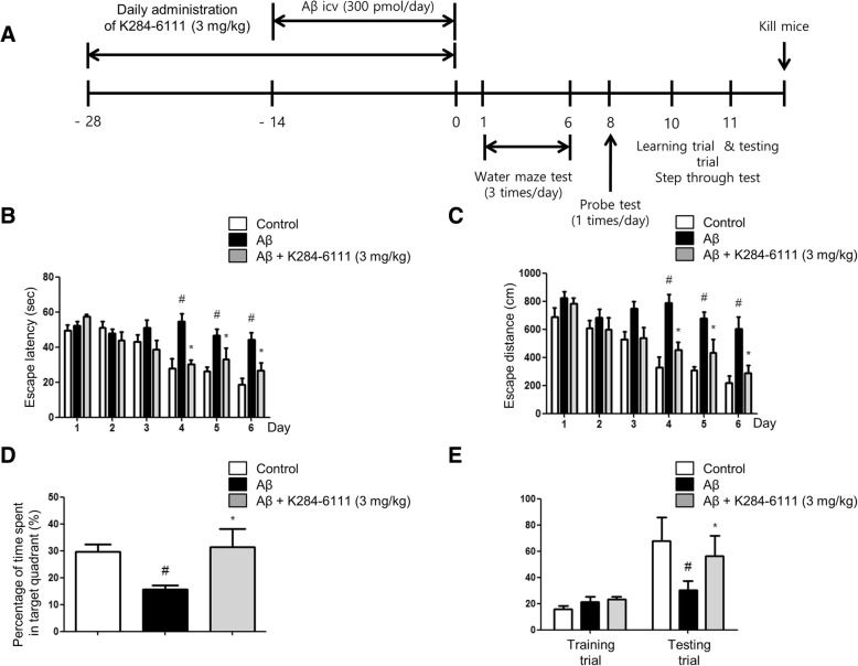
Methods: We examined whether K284-6111 (3 mg/kg given orally for 4 weeks) prevents amyloidogenesis and memory loss in Aβ1-42-induced AD mice model. After intracerebroventrical (ICV) infusion of Aβ1-42 for 14 days, the cognitive function was assessed by the Morris water maze test and passive avoidance test. K284-6111 treatment was found to reduce Aβ1-42-induced memory loss.

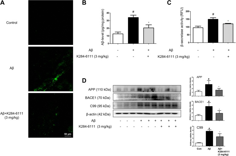
Results: A memory recovery effect was found to be associated with the reduction of Aβ1-42-induced expression of inflammatory proteins (iNOS, COX-2, GFAP, and Iba-1) and the suppression of CHI3L1 expression in the brain. Additionally, K284-6111 reduced Aβ1-42-induced β-secretase activity and Aβ generation. Lipopolysaccharide (LPS)-induced (1 μg/mL) expression of inflammatory (COX-2, iNOS, GFAP, Iba-1) and amyloidogenic proteins (APP, BACE1) were decreased in microglial BV-2 cells and cultured astrocytes by the K284-6111 treatment (0.5, 1, and 2 μM). Moreover, K284-6111 treatment suppressed p50 and p65 translocation into the nucleus, and phosphorylation of IκB in vivo and in vitro.

Conclusion: These results suggest that CHI3L1 inhibitor could be an applicable intervention drug in amyloidogenesis and neuroinflammation, thereby preventing memory dysfunction via inhibition of NF-κB.

Keywords: Alzheimer’s disease; Amyloidogenesis; CHI3L1; NF-κB; Neuroinflammation.


Conflict of interest statement

The experimental protocols were carried out according to the guidelines for animal experiments of the Institutional Animal Care and Use Committee (IACUC) of Laboratory Animal Research Center at Chungbuk National University, Korea (CBNUA-1073-17-01).

Not applicable.

The authors declare that they have no competing interests.

Springer Nature remains neutral with regard to jurisdictional claims in published maps and institutional affiliations.

Figures

Fig. 1
Fig. 1
K284-6111 improves memory impairment in Aβ1–42-infused mice. a Timeline depicts the treatment of K284-6111 and assessments of the cognitive functions of mice. The male mice (n = 10) were orally treated with K284-6111 at a daily dose of 3 mg/kg for 4 weeks. After infusion of Aβ1–42 (300 pmol/day) for 14 days, memory tests were conducted. The training trial was performed two times a day for 7 days. Swimming time (b) and swimming distance (c) to the platform were automatically recorded. The time spent in the target quadrant and target site crossing within 60 s were represented (d). To perform the passive avoidance test, step-through method was used (e). Each value is presented as mean ± S.D. from 10 male mice. #Significant difference to control mice (P < 0.05), *Significant difference to Aβ1–42-injected mice (P < 0.05)
Fig. 2
Fig. 2
K284-6111 on Aβ1–42-infused mice in both GFAP and IBA-1-positive mice brain. Immunostaining of CHI3L1, iNOS, GFAP, and Iba-1 proteins in the hippocampus were performed in 20-μm-thick sections of mice brain with specific primary antibodies and biotinylated secondary antibodies (a, b). The expression of CHI3L1, GFAP, Iba-1, COX-2, and iNOS were detected by Western blotting using specific antibodies in the mice brain. For the cropped images, samples were run in the same gels under the same experimental conditions and processed in parallel. Band density was quantified from three mice (c). mRNA levels of CHI3L1 (d), TNF-α (e), IL-1β (f), and IL-6 (g) were detected by qRT-PCR in hippocampus. #Significant difference to control mice (P < 0.05), *Significant difference to Aβ1–42-injected mice (P < 0.05)
Fig. 3
Fig. 3
K284-6111 inhibits accumulation of Aβ1–42 in the brain of Aβ1–42-infused mice brain. a Aβ accumulation in the brains of Aβ1–42-infused mice was determined by thioflavin S staining. The levels of Aβ1–42 in mice brain (n = 5) were measured by ELISA (b). The activity of β-secretase in mice brain (n = 5) was investigated using an assay kit (c). The expression of APP, BACE1, and C99 were detected by Western blotting using specific antibodies in the mice brain (d). For the cropped images, samples were run in the same gels under the same experimental conditions and processed in parallel. Band density was quantified from three mice (d). #Significant difference to control mice (P < 0.05), *Significant difference to Aβ1–42-injected mice (P < 0.05)
Fig. 4
Fig. 4
K284-6111 on NF-κB translocation related DNA-binding activity-mediated CHI3L1. Phosphorylation of IκB, and p50 and p65 translocation was detected by Western blotting using specific antibodies in mice brain. β-actin and HistoneH1 protein was used as an internal control, and the graphs represented the arbitrary density of blot signal (a). Band density was quantified from three mice (a). Whole cell lysates of B16F10 were incubated with K284-6111-conjugated Sepharose 4B. After precipitation, the levels of bound CHI3L1 were monitored by Western blot analysis (b). Docking model of K284-6111 (c). CHI3L1 siRNA augmented K284-6111-induced inhibitory effect on NF-κB expression and inflammation (d). Each value is mean ± S.D. from three experiments (d). *Significant difference from control group (p < 0.05). # Significant difference from Aβ1–42-infused group (p < 0.05)
Fig. 5
Fig. 5
K284-6111 inhibits NF-κB translocation in microglial BV-2 cells and cultured astrocytes. The cultured microglial BV-2 cells (a) and cultured astrocytes (b) were incubated with anti-p65 (red) and DAPI staining (blue). Phosphorylation of IκB, and p50 and p65 translocation were detected by Western blotting using specific antibodies in microglial BV-2 cells (c) and cultured astrocytes (d). Each value under the bands is mean ± S.D. from three experiments (c, d). #Significantly different from control group (p < 0.05). *Significantly different from LPS-treated group (p < 0.05)
Fig. 6
Fig. 6
K284-6111 on neuroinflammatory and amyloidogenic responses in microglial BV-2 cells and astrocytes. NO level was measured in K284-6111-treated microglial BV-2 cells (a) and astrocytes (b). CHI3L1, iNOS, COX-2, and Iba-1 proteins were detected by Western blotting using specific antibodies in K284-6111-treated microglial BV-2 cells (c). CHI3L1, iNOS, COX-2, and GFAP proteins were detected by Western blotting using specific antibodies in K284-6111-treated cultured astrocytes (d). APP and BACE1 proteins were detected by Western blotting using specific antibodies in K284-6111-treated microglial BV-2 cells (e) and cultured astrocytes (f). Each value under the band is mean ± S.D. from three experiments (c, d, e, f). #Significantly different from control group (p < 0.05). *Significantly different from LPS-treated group (p < 0.05)

References

    1. Alzheimer's A. Alzheimer’s disease facts and figures. Alzheimers Dement. 2016;2016(12):459–509. - PubMed
    1. Fraser PE, Yang DS, Yu G, Levesque L, Nishimura M, Arawaka S, Serpell LC, Rogaeva E, St George-Hyslop P. Presenilin structure, function and role in Alzheimer disease. Biochim Biophys Acta. 2000;1502:1–15. doi: 10.1016/S0925-4439(00)00028-4. - DOI - PubMed
    1. Takahashi RH, Nagao T, Gouras GK. Plaque formation and the intraneuronal accumulation of beta-amyloid in Alzheimer's disease. Pathol Int. 2017;67:185–193. doi: 10.1111/pin.12520. - DOI - PubMed
    1. Hatami A, Monjazeb S, Milton S, Glabe CG. Familial Alzheimer’s disease mutations within the amyloid precursor protein alter the aggregation and conformation of the amyloid-beta peptide. J Biol Chem. 2017;292:3172–3185. doi: 10.1074/jbc.M116.755264. - DOI - PMC - PubMed
    1. Lasagna-Reeves CA, Castillo-Carranza DL, Sengupta U, Clos AL, Jackson GR, Kayed R. Tau oligomers impair memory and induce synaptic and mitochondrial dysfunction in wild-type mice. Mol Neurodegener. 2011;6:39. doi: 10.1186/1750-1326-6-39. - DOI - PMC - PubMed

Publication types

MeSH terms