Fatty pancreas: A possible risk factor for pancreatic cancer in animals and humans
- PMID: 30099827
- PMCID: PMC6172058
- DOI: 10.1111/cas.13766
Fatty pancreas: A possible risk factor for pancreatic cancer in animals and humans
Abstract
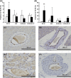
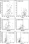
Obesity, type 2 diabetes mellitus (T2DM) and aging are associated with pancreatic cancer risk, but the mechanisms of pancreatic cancer development caused by these factors are not clearly understood. Syrian golden hamsters are susceptible to N-nitrosobis(2-oxopropyl)amine (BOP)-induced pancreatic carcinogenesis. Aging, BOP treatment and/or a high-fat diet cause severe and scattered fatty infiltration (FI) of the pancreas with abnormal adipokine production and promote pancreatic ductal adenocarcinoma (PDAC) development. The KK-Ay mouse, a T2DM model, also develops severe and scattered FI of the pancreas. Treatment with BOP induced significantly higher cell proliferation in the pancreatic ducts of KK-Ay mice, but not in those of ICR and C57BL/6J mice, both of which are characterized by an absence of scattered FI. Thus, we hypothesized that severely scattered FI may be involved in the susceptibility to PDAC development. Indeed, severe pancreatic FI, or fatty pancreas, is observed in humans and is associated with age, body mass index (BMI) and DM, which are risk factors for pancreatic cancer. We analyzed the degree of FI in the non-cancerous parts of PDAC and non-PDAC patients who had undergone pancreatoduodenectomy by histopathology and demonstrated that the degree of pancreatic FI in PDAC cases is significantly higher than that in non-PDAC controls. Moreover, the association with PDAC is positive, even after adjusting for BMI and the prevalence of DM. Accumulating evidence suggests that pancreatic FI is involved in PDAC development in animals and humans, and further investigations to clarify the genetic and environmental factors that cause pancreatic FI are warranted.
Keywords: cancer susceptibility; fatty infiltration; obesity; pancreatic cancer; type 2 diabetes mellitus.
© 2018 The Authors. Cancer Science published by John Wiley & Sons Australia, Ltd on behalf of Japanese Cancer Association.
Figures


References
-
- Cancer Statistics in Japan . Foundation for Promotion of Cancer Research. Tokyo, Japan: Cancer Statistics in Japan; 2016.
-
- Hori M, Kitahashi T, Imai T, et al. Enhancement of carcinogenesis and fatty infiltration in the pancreas in N‐nitrosobis(2‐oxopropyl)amine‐treated hamsters by high‐fat diet. Pancreas. 2011;40:1234‐1240. - PubMed
-
- Takahashi M, Kitahashi T, Ishigamori R, et al. Increased expression of inducible nitric oxide synthase (iNOS) in N‐nitrosobis(2‐oxopropyl)amine‐induced hamster pancreatic carcinogenesis and prevention of cancer development by ONO‐1714, an iNOS inhibitor. Carcinogenesis. 2008;29:1608‐1613. - PubMed
-
- Takeuchi Y, Takahashi M, Sakano K, et al. Suppression of N‐nitrosobis(2‐oxopropyl)amine‐induced pancreatic carcinogenesis in hamsters by pioglitazone, a ligand of peroxisome proliferator‐activated receptor γ. Carcinogenesis. 2007;28:1692‐1696. - PubMed
-
- Pour PM, Lawson T. Pancreatic carcinogenic nitrosamines in Syrian hamsters. IARC Sci Publ. 1984;57:683‐688. - PubMed
Publication types
MeSH terms
Substances
Grants and funding
- 15K14393/Grant-in-Aid for Scientific Research from the Japan Society for the Promotion of Science
- 18K07081/Grant-in-Aid for Scientific Research from the Japan Society for the Promotion of Science
- 25290049/Grant-in-Aid for Scientific Research from the Japan Society for the Promotion of Science
- Research Grant of the Princess Takamatsu Cancer Research Fund
- grant of the Third-Term Comprehensive 10-Year Strategy for Cancer Control from the Ministry of Health, Labor, and Welfare of Japan
- Grants-in-Aid from the Foundation for Promotion of Cancer Research and the Pancreas Research Foundation of Japan
- 21-2-1/Grants-in-Aid for Cancer Research from the Ministry of Health, Labour, and Welfare of Japan and Management Expenses Grants from the Government to the National Cancer Center
- National Cancer Center Research Core Facility
LinkOut - more resources
Full Text Sources
Other Literature Sources
Medical

