. 2018 Aug 23;174(5):1106-1116.e9.
doi: 10.1016/j.cell.2018.06.038. Epub 2018 Aug 9.

Crystal Structure of the COMPASS H3K4 Methyltransferase Catalytic Module

Affiliations

Crystal Structure of the COMPASS H3K4 Methyltransferase Catalytic Module

Peter L Hsu et al. Cell. .

Abstract

The SET1/MLL family of histone methyltransferases is conserved in eukaryotes and regulates transcription by catalyzing histone H3K4 mono-, di-, and tri-methylation. These enzymes form a common five-subunit catalytic core whose assembly is critical for their basal and regulated enzymatic activities through unknown mechanisms. Here, we present the crystal structure of the intact yeast COMPASS histone methyltransferase catalytic module consisting of Swd1, Swd3, Bre2, Sdc1, and Set1. The complex is organized by Swd1, whose conserved C-terminal tail not only nucleates Swd3 and a Bre2-Sdc1 subcomplex, but also joins Set1 to construct a regulatory pocket next to the catalytic site. This inter-subunit pocket is targeted by a previously unrecognized enzyme-modulating motif in Swd3 and features a doorstop-style mechanism dictating substrate selectivity among SET1/MLL family members. By spatially mapping the functional components of COMPASS, our results provide a structural framework for understanding the multifaceted functions and regulation of the H3K4 methyltransferase family.

Keywords: COMPASS; H3K4 methylation; MLL; Set1; chromatin; epigenetics; methyltransferases; structural biology.


Conflict of interest statement

DECLARATION OF INTERESTS

The authors declare no competing interests.

Figures
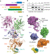
Figure 1.
Figure 1.. Overall architecture of the yeast COMPASS catalytic module
(A) Domain organization and construct design of yeast Set1, Swd1, Bre2, Swd3, and Sdc1. Only the SET domain of Set1 is shown. (B) Time course of H3K4 methylation catalyzed by purified yeast COMPASS catalytic module with recombinant mono-nucleosome substrate. Reactions were quenched at specified time points, and analyzed by SDS-PAGE, followed by western blotting using methylation specific antibodies. Time point 0 min was taken seconds after addition of enzyme to the reaction. (C) Overall structure of the COMPASS catalytic module. Set1 (blue), Swd1 (magenta), Swd3 (green), Bre2 (orange), and two copies of Sdc1 (yellow and turquoise) are shown in cartoon form. Both the H3 peptide (white) and SAM cofactor (yellow) are shown in space filling model. The long C-terminal tail of Swd1 and unique SMART loop of Swd3 are shown in tube representation to highlight their positions in the complex. (D) Individual subunits of the catalytic module are shown in surface representation to illustrate their relative spatial relationships. Orientation of the subunits is the same as shown in (C). For Swd1, from left to right, arrows show the winding path of the C-terminal tail. A dashed line in the tail illustrates a disordered region not observed in the structure.
Figure 2.
Figure 2.. Bre2/ASH2L fold into an extended non-canonical SPRY domain
(A) Crystal structure of human ASH2L (grey) bound to a peptide of RBBP5 (magenta) (PDB: 4X8P). Dashed lines indicate the absent N-, insertion, and C-terminal sequences in the structure. (B) Structure of yeast Bre2. The conserved SPRY-only domain and Ins-2 is shown in orange, while the N-terminal pre-SPRY and Ins-1 are shown in blue and pale pink, respectively. The C-terminal helix is colored in green. Small arrowheads indicate the terminal ends of the insertion elements. The Swd1 peptide equivalent to that of RBBP5 in (A) is shown in purple. Structure is depicted in smooth lines representation for clarity. (C) Surface representation of Set1 (blue) and Bre2 (orange). A highly conserved surface formed by the auxiliary β-sheet in Bre2 is highlighted in red. (D) View of Bre2 looking down the C-terminal helix (green), illustrating the position of the auxiliary β-sheet right above the helix. Sdc1-A (yellow) and Sdc1-B (turquoise) prop up this sheet.
Figure 3.
Figure 3.. Sdc1 assembles as an asymmetric dimer to bind Bre2
(A) Left panel: structure of DPY-30 (turquoise and yellow) bound to the C-terminal helix of ASH2L (green) (PDB: 4RIQ). Right panel: equivalent view of Sdc1 (yellow and turquoise) bound to the Bre2 C-terminal helix (green). A dashed line in Sdc1-B denotes a poorly structured loop that was not clearly resolved in the crystal. (B) Superposition of the two protomers of Sdc1 highlighting the two different positions of the same R88 side chain in the individual copies. (C) Close-up view of the interface between Sdc1-A (turquoise) and Bre2 (orange and green). R88 of Sdc1 forms a salt bridge with Bre2-D401 on the C-terminal helix and a backbone hydrogen bonding interaction with the body of Bre2. (D) Close-up view of the interface between Sdc1-B (yellow) and Bre2 (orange and green). The same R88 seen in (C) is facing towards Sdc1-A with the rest of the N-terminus packing against Bre2 near the auxiliary β-sheet. (E) Glutathione affinity co-purification of GST-Sdc1 N-terminal truncation mutants and His-Bre2 co-expressed in insect cell. Protein interactions were assessed by SDS-PAGE followed by Coomassie staining. Inputs from soluble lysates were detected using antibodies against the respective tags.
Figure 4.
Figure 4.. The C-terminal tail of Swd1 organizes the catalytic module and forms a key pocket with Set1
(A) A global view of the catalytic module highlighting the path of the Swd1 C-terminal tail. Set1 (blue), Swd3 (green), and Bre2 (orange) are shown in surface representation, while Swd1 (magenta) and its C-terminal tail (pink) are shown in cartoon form. A dotted line connecting the two halves of the C-terminal tail represents a disordered region not seen in the structure. (B) Close-up view of the interface between Swd1-WDRP loop (pink) with Swd3 (green) and Set1 (blue). Swd3 and Set1 are shown in surface representation, while the WDRP region is shown in cartoon. Highly conserved Swd1 residues involved in interacting with both Set1 and the Swd3 loop are shown in sticks. The two sub-domains of Set1 are labeled as SET-N/C and SET-I. (C) Alternate view of the Set1-Swd1-Swd3 interface highlighting the crevasse formed between Set1 (blue) and Swd1-WDRP loop (pink), both of which are shown in surface representation. The residues from Swd1 that line this pocket are labeled. The Swd3 SMART loop (green) shown in cartoon inserts a tryptophan residue (sticks) into the Set1-Swd1 pocket. The SAM cofactor and the H3 peptide are shown in CPK form and sticks, respectively. (D) Path of the Swd1 distal tail that recruits Swd3 to the catalytic module. The tail is shown in cartoon form in pink, while the WD40 domains of Swd3 and Swd1 are shown in surface representation in green and magenta, respectively. (E) Glutathione affinity co-purification of full-length (FL) GST-Swd1 and N-terminal (ΔN) and C-terminal truncation (ΔC) mutants with His-Swd3 co-expressed in insect cells. Protein interactions were assessed by SDS-PAGE followed by Coomassie staining. His-Swd3 input from soluble lysates were detected using antibodies against the His-tag.
Figure 5.
Figure 5.. The SMART loop of Swd3 regulates Set1 activity through the Kabuki pocket
(A) Close up view of the Set1-Swd1-Swd3 interface, centered around the Swd3 SMART loop. Set1 and Swd1 (blue and pink respectively) are shown in surface, while the Swd3 loop is in cartoon form. The residues that form the SMART loop are shown in sticks. Dashed lines indicate hydrogen bonds. (B) Detailed view of the residues that form the Kabuki pocket. On the Set1 side of the pocket are the amino acids W870, I909, Y913, T926, R933 and F934, while the cofactor SAM sits at the periphery. Swd1 contributes W356, L359, D362, and F363 to the formation of the pocket. The Swd3 tryptophan residue, W197, inserted into the pocket is shown in sticks without the rest of the Swd3 SMART loop. (C) H3K4 methyltransferase activities of yeast and mammalian catalytic module containing wild type and mutant Swd3 and two different constructs of SETD1B against nucleosomes. Left panel shows the yeast catalytic module with Swd3 bearing a W197A mutation. Right panel shows the activity of the assembled SETD1B catalytic module containing the SETD1B C-terminal WIN-SET domain excluding (-DWLND) or including (+DWLND) the putative SMART motif shown in (D). (D) Sequence alignment of mammalian SETD1A and SETD1B. The putative SMART motif of mammalian SETD1A/SETD1B is upstream of the conserved WDR5-interacting motif (WIN). For clarity, the SET domain of each SETD1A/B vertebrate ortholog C-terminal to the WIN motif is not shown. The SMART motif from K. lactis Swd3 is shown below the mammalian sequence as a comparison. A gradient of red to blue colors are used to indicate low to high degree of conservation.
Figure 6.
Figure 6.. A “doorstop” mechanism determines substrate specificity in SET1/MLL enzymes
(A) Superposition of yeast Set1 (blue), MLL3 (orange, PDB: 5F6K chain C), and MLL1 (purple, PDB: 5F6I). Helices are shown as cylinders for clarity. Note the variability in the SET-I helix between the three structures superposed, shifting between an open and closed state. The side chain of the Arg933 residue at the bottom of the Kabuki pocket is shown in sticks as a reference point for orientation. (B) View looking down the Kabuki pocket, highlighting the door loop of yeast Set1 (blue). The side chains of the door loop are shown as sticks. Residues that line the Kabuki pocket from the Swd1 side are shown in pink sticks. SAM and the H3 peptide are shown in yellow and grey sticks to illustrate the spatial relationship of the door loop to both the cofactor and substrate. (C) Sequence alignment of yeast Set1 and human SET1/MLL family members surrounding the SET-I helix and the following door loop. (D) H3K4 methyltransferase activity of yeast and mammalian catalytic module containing Set1 and MLL3 door loop mutants against nucleosome substrates. Left panel are Set1 mutants with mutations in the second half of the door loop motif showing gradual reduction of tri-methylation upon introduction of bulkier side chains. Left panel are mutants of MLL3-WRAD with mutations in the doorstop position of the loop. The simple replacement of NR with GI results in robust gain of function for MLL3. (E) Superposition of MLL3 in its closed conformation (orange, PDB: 5F6K chain C) with Set1 (blue) bound to Swd1 (pink). Arg4822 on MLL3 forms a perfect salt bridge with D362 of Swd1, whose corresponding residue in RBBP5 is D335. Doorstop in green indicates a geometrically allowed structural configuration. (F) Superposition modeling of RBBP5/Swd1-bound MLL3 (orange, PDB: 5F6K chain E) in the experimentally determined open state and MLL1 (purple, PDB: 5F6I) in the open state based on the yeast catalytic module structure. Arg4822 of MLL3 clashes with Asp362 on Swd1, while Ile3880 of MLL1 can fully open even in the presence of Swd1/RBBP5. Doorstop in red indicates a geometrically disallowed structural configuration. (G) H3K4 methyltransferase activity of the mammalian MLL3-centered catalytic module containing wild type (WT) RBBP5 and a RBBP5-D335A mutant against nucleosome substrates. The catalytic module with the RBBP5 mutant produced an increased amount of di-and tri-methylated H3K4 products relative to wild-type protein.

Similar articles

Cited by

References

    1. Adams PD, Afonine PV, Bunkóczi G, Chen VB, Davis IW, Echols N, Headd JJ, Hung L-W, Kapral GJ, Grosse-Kunstleve RW, et al. (2010). PHENIX: a comprehensive Python-based system for macromolecular structure solution. Acta Crystallogr. D Biol. Crystallogr 66, 213–221. - PMC - PubMed
    1. Allis CD, Caparros M-L, Jenuwein T, and Reinberg D (2015). Epigenetics (Cold Spring Harbor Laboratory Press; ).
    1. Ardehali MB, Mei A, Zobeck KL, Caron M, Lis JT, and Kusch T (2011). Drosophila Set1 is the major histone H3 lysine 4 trimethyltransferase with role in transcription. EMBO J 30, 2817–2828. - PMC - PubMed
    1. Avdic V, Zhang P, Lanouette S, Groulx A, Tremblay V, Brunzelle J, and Couture J-F (2011). Structural and Biochemical Insights into MLL1 Core Complex Assembly. Structure 19, 101–108. - PubMed
    1. Briggs SD, Xiao T, Sun Z-W, Caldwell JA, Shabanowitz J, Hunt DF, Allis CD, and Strahl BD (2002). Gene silencing: Trans-histone regulatory pathway in chromatin. Nature 418, 498. - PubMed

Publication types

MeSH terms

LinkOut - more resources