. 2018 Oct:16:160-171.
doi: 10.1016/j.molmet.2018.07.008. Epub 2018 Jul 27.

TGF-β receptor 1 regulates progenitors that promote browning of white fat

Affiliations

TGF-β receptor 1 regulates progenitors that promote browning of white fat

Umesh D Wankhade et al. Mol Metab. 2018 Oct.

Abstract

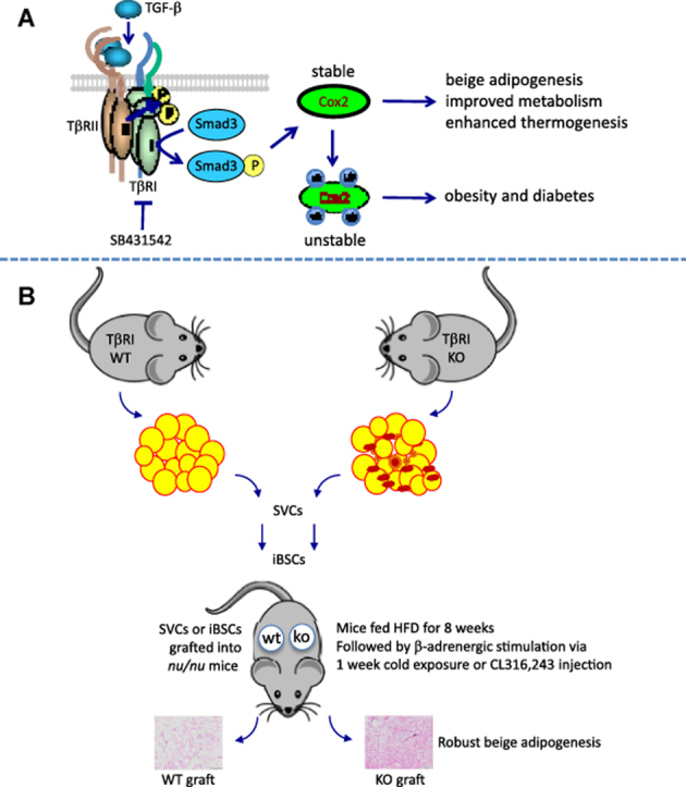
Objective: Beige/brite adipose tissue displays morphological characteristics and beneficial metabolic traits of brown adipose tissue. Previously, we showed that TGF-β signaling regulates the browning of white adipose tissue. Here, we inquired whether TGF-β signals regulated presumptive beige progenitors in white fat and investigated the TGF-β regulated mechanisms involved in beige adipogenesis.

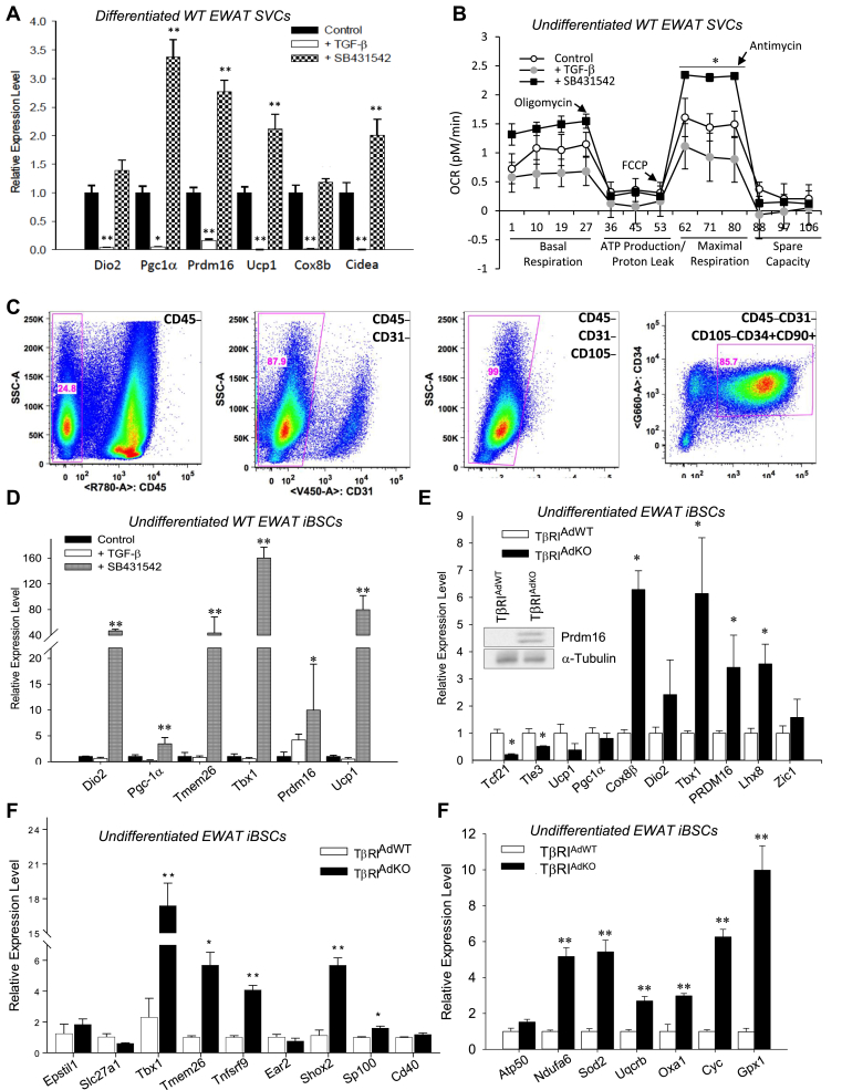
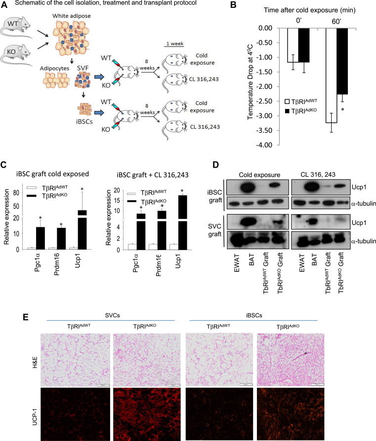
Methods: We deleted TGF-β receptor 1 (TβRI) in adipose tissue (TβRIAdKO mice) and, using flow-cytometry based assays, identified and isolated presumptive beige progenitors located in the stromal vascular cells of white fat. These cells were molecularly characterized to examine beige/brown marker expression and to investigate TGF-β dependent mechanisms. Further, the cells were transplanted into athymic nude mice to examine their adipogenesis potential.

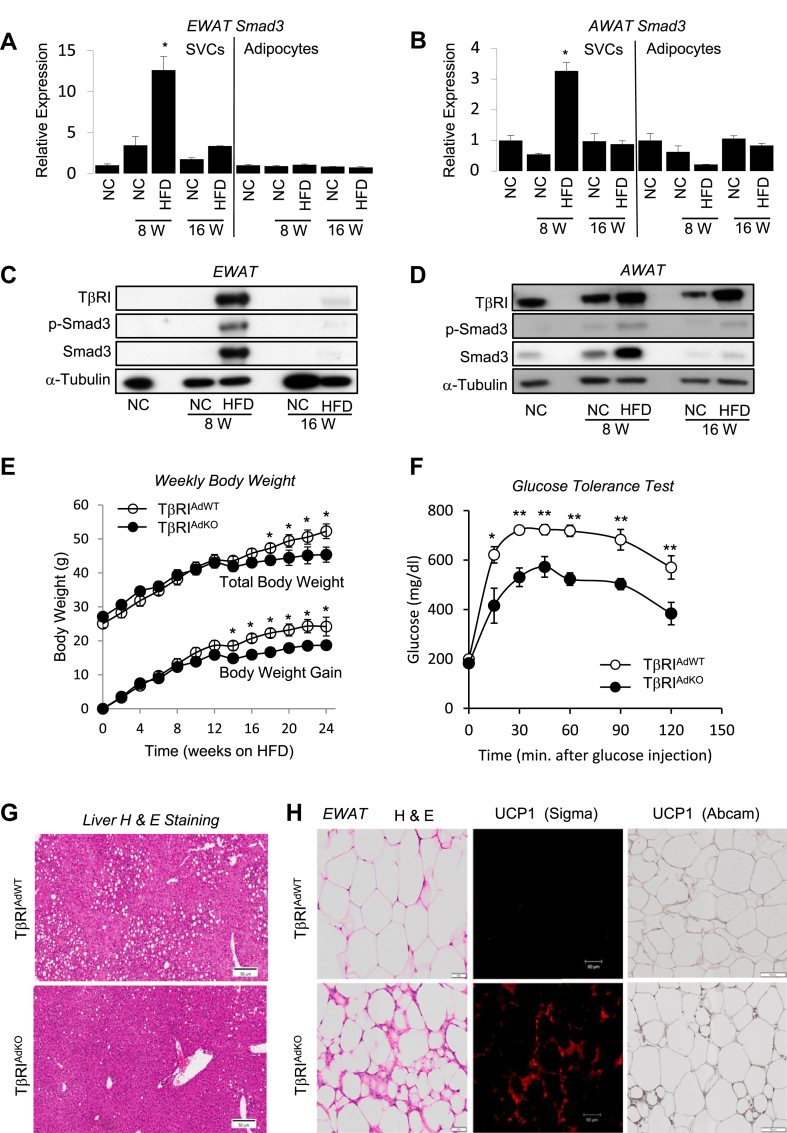
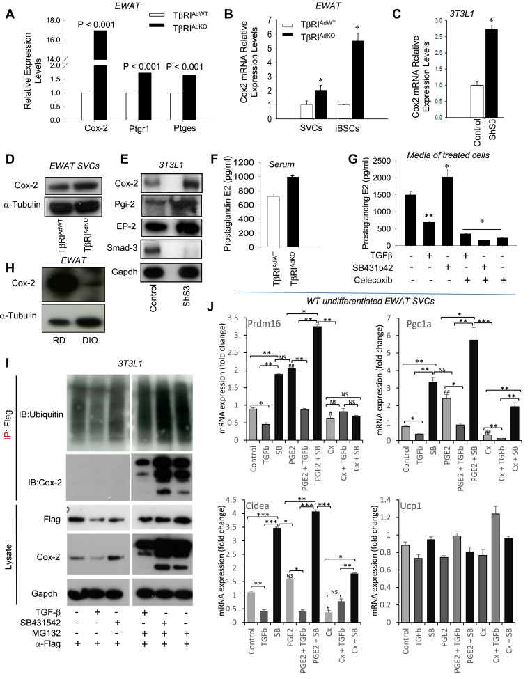
Results: Deletion of TβRI promotes beige adipogenesis while reducing the detrimental effects of high fat diet feeding. Interaction of TGF-β signaling with the prostaglandin pathway regulated the appearance of beige adipocytes in white fat. Using flow cytometry techniques and stromal vascular fraction from white fat, we isolated presumptive beige stem/progenitor cells (iBSCs). Upon genetic or pharmacologic inhibition of TGF-β signaling, these cells express high levels of predominantly beige markers. Transplantation of TβRI-deficient stromal vascular cells or iBSCs into athymic nude mice followed by high fat diet feeding and stimulation of β-adrenergic signaling via CL316,243 injection or cold exposure promoted robust beige adipogenesis in vivo.

Conclusions: TβRI signals target the prostaglandin network to regulate presumptive beige progenitors in white fat capable of developing into beige adipocytes with functional attributes. Controlled inhibition of TβRI signaling and concomitant PGE2 stimulation has the potential to promote beige adipogenesis and improve metabolism.

Keywords: Beige/brite adipogenesis; Cyclooxygenase 2; Diabetes; Metabolism; Obesity; Progenitors; Prostaglandin E2; TGF-beta.


Figures

Image 1
Graphical abstract
Figure 1
Figure 1
Adipose tissue specific deletion of TGFβ receptor 1 promotes browning of white fat and protects from obesity, glucose intolerance and hepatic steatosis. (A-D) Normal mice (6-week-old, males, n = 4) were fed either normal chow diet (NC) or HFD for 8 weeks or 16 weeks and SVCs and adipocytes from the EWAT (A, C) and AWAT (B, D) were examined for activation of TGF-β/Smad3 signaling. Increase in Smad3 transcripts (A, B) was seen in SVCs isolated from EWAT (A) and AWAT (B), but not in mature adipocytes, in response to 8 weeks of HFD feeding. Increased levels of phosphorylated Smad3 protein was also seen in the SVC fraction of EWAT (C) and AWAT (D) in response to 8 weeks of HFD feeding. Levels of TβRI were also increased under those conditions (C and D). Total Smad3 and loading control Tubulin protein are shown. (E–G) HFD fed TβRIAdKO mice (male, n = 6), compared to similarly fed age-matched TβRIAdWT mice (male, n = 9) exhibit reduced weight gain (E), improved glucose tolerance (F) and are protected from hepatic steatosis (G). (H) EWAT depots from HFD fed TβRIAdKO mice exhibit smaller sized multilocular adipocytes and increased UCP1 positive cells as detected via immunofluorescence (using Sigma antibody) and immunohistochemistry (using Abcam antibody). T-test, one-way ANOVA and group wise differences were assessed by post-hoc Tukey HSD test wherever appropriate. Error Bars are expressed as ± Standard Errors, *p < 0.05; **p < 0.005; ***p < 0.001.
Figure 2
Figure 2
TGF-β signaling regulates inducible beige progenitor cells. (A) TGF-β represses whereas the TGF-β receptor 1 inhibitor SB431542 stimulates the expression of brown/beige marker genes in normal wild type SVCs differentiated using brown adipocyte differentiation cocktail. SVCs were isolated from six-week aged male mice fed HFD for 14–16 weeks. Levels shown in TGF-β (open columns) and SB431542 (shaded columns) treated samples are statistically compared to Control (closed columns) treated samples. (n = 3 each) (B) Treatment of wild type undifferentiated SVCs with TGFβ leads to reduced basal and maximal respiration, whereas SB431542 treatment reverses the effect and leads to significantly higher respiration capacity. SVCs were treated with oligomycin (0.5 μM), FCCP (1 μM) and antimycin A (0.25 μM) as described in the methods section. (n = 3 each) (C) SVCs were sorted using flow cytometry and specific cell surface antibodies to identify putative beige stem cells. The boxed regions within the panels indicate the percentage of cells sorted using specific antibodies as identified in the top right corner of the panel. (n = 3 each) (D) SB431542, but not TGF-β, stimulates the expression of brown/beige marker genes (Dio2, Pgc-1α, Tmem26, Tbx1, Prdm16, and Ucp1) in normal undifferentiated wild type iBSCs. Levels shown in TGF-β (open columns) and SB431542 (shaded columns) treated samples are statistically compared to Control (closed columns) treated samples. (n = 3 each). (E) Compared to undifferentiated iBSCs isolated from TβRIAdWT adipose tissue, the undifferentiated TβRIAdKO iBSCs exhibit increased expression of brown/beige specific markers Tbx1, Prdm16, Lhx8, and Cox8b. WAT specific markers Tcf21 and Tle3 are repressed. Brown specific Zic1 transcript and levels of functional brown/beige markers Ucp1, Dio2, and Pgc-1α are not changed. Epididymal adipose tissue depots were collected and pooled from n = 3 mice for iBSC isolation. Levels of Prdm16 protein are elevated in TβRIAdKO iBSCs (see inset); equal tubulin protein levels are shown as protein loading control. (F) Undifferentiated TβRIAdKO iBSCs exhibit increased expression of nine beige specific markers. Epididymal adipose tissue depots were collected and pooled from n = 3 mice of each genotype for iBSC isolation. (G) Compared to undifferentiated TβRIAdWT iBSCs, the undifferentiated TβRIAdKO iBSCs exhibit increased expression of mitochondrial specific markers. Data are analyzed by one-way ANOVA and group wise differences were assessed by post-hoc Tukey HSD test wherever appropriate. Error Bars are expressed as ± Standard Errors, *p < 0.05; **p < 0.005; ***p < 0.001.
Figure 3
Figure 3
TGF-β/Smad3 signaling regulates proteasomal degradation of Cox-2 and interaction between TGF-β and prostaglandin pathways regulate beige marker genes. (A) Ptgs2/Cox-2 gene expression is increased 17-fold in HFD-fed EWAT tissues from TβRIAdKO mice. (B, C) SVCs and iBSCs from TβRIAdKO mice (B) and 3T3L1-ShS3 cells (C) exhibit increased expression of Cox-2 mRNA. (D, E) Increased protein expression of Cox-2 and prostaglandin pathway intermediates (Pgi-2, EP-2) in TβRIAdKO EWAT tissue (D) and in 3T3L1-shSmad3 cells (E). α-tubulin and GAPDH levels are shown as loading control. Smad3 protein expression is observed in control 3T3L1 cells, but not in 3T3L1-shSmad3 cells. (F) Levels of prostaglandin E2 are elevated in cell culture media of TβRIAdKO iBSCs (n = 3 each treatment). (G) TGF-β treatment suppressed, whereas SB431542 increased, prostaglandin E2 levels in control wild-type SVCs. Treatment with the Cox-2 inhibitor Celecoxib suppressed prostaglandin E2 levels. Treated samples are statistically compared to vehicle-treated samples (first column) (n = 3 each treatment). (H) Reduced Cox-2 protein level in diet-induced obese (DIO) mice. (I) Control 3T3L1 cells were transfected with Flag-tagged ubiquitin plasmids followed by treatment with TGFβ or SB431542 in the presence or absence of MG132. Cells were harvested for protein followed by immunoblot of the lysates to detect relative expression of Flag-tagged ubiquitin, Cox-2 and GAPDH control (lower three panels). Additionally, equal amounts of lysates were subjected to immunoprecipitation with anti-Flag antibodies followed by immunoblot with anti-Cox-2 antibodies to detect ubiquitinated Cox-2 in cells treated with TGF-β or SB431542 with or without the presence of MG132 (upper two panels). (J) Interaction between the TGF-β and prostaglandin pathways on Pgc-1α, Cidea, Prdm16, and Ucp1 gene expression. SVCs were isolated from EWAT of WT mice and cultured with ligands TGF-β1 (5 ng/ml) and PGE2 (10 ug/ml) or inhibitors SB431542 (10 uM) and Celecoxib (Cx) (10 uM) either singly or in combination as outlined in the figure for 16 h after which the cells were harvested and analyzed for gene expression (n = 3 each per treatment). Data are analyzed by one-way ANOVA and group wise differences were assessed by post-hoc Tukey HSD test wherever appropriate. Error Bars are expressed as ± Standard Errors, *p < 0.05; **p < 0.005; ***p < 0.001 (among the groups indicated) and p values #p < 0.05, ##p < 0.01 ###p < 0.001 for values compared between control vs PGE2 and Cx groups. NS represents not-significant.
Figure 4
Figure 4
TβRIAdKO iBSCs undergo functional beige adipogenesis in vivo. (A) Schematic representation of the transplant protocol. iBSCs and SVCs isolated from TβRIAdWT and TβRIAdKO adipose tissue were transplanted into the flanks of athymic nu/nu mice (n = 6 mice, 3 per treatment i.e. CL316,243 and cold exposure). FACS sorted cells isolated from TβRIAdWT and TβRIAdKO mice were transplanted into opposite flanks as shown to allow comparison of graft development within the same recipient mouse. Upon transplantation, the mice were fed HFD for eight weeks after which they were either subjected to 4 °C cold exposure (90 min/day for seven consecutive days) or injected with the β-adrenergic receptor agonist CL316,243 (1 mg/kg bw per day for seven consecutive days). (B) During cold exposure, the local surface temperature of the TβRIAdKO iBSCs graft was significantly warmer compared to the TβRIAdWT iBSCs graft. (C) Transcript levels of Ucp1, Prdm16 and Pgc-1α were increased in TβRIAdKO iBSCs grafts upon cold exposure and stimulation by CL316,243. (D) UCP1 protein expression was highly elevated in TβRIAdKO iBSC and SVC grafts, compared to that seen in the TβRIAdWT iBSC graft. Levels of UCP1 protein seen in the TβRIAdKO iBSC graft were comparable to levels of UCP1 seen in the BAT harvested from the same recipient mouse. In comparison, EWAT shows undetectable UCP1 expression. (E) Analysis of the grafts following the 8-week growth and 1-week β-adrenergic receptor pathway stimulation. Compared to grafts from TβRIAdWT donors, H&E staining and UCP1 immune staining of the SVC and iBSC grafts from TβRIAdKO donors exhibited abundant UCP1 positive small-sized multilocular adipocytes. Data are analyzed by T-test. Error Bars are expressed as ± Standard Errors, *p < 0.05.

Similar articles

Cited by

References

    1. Abel E.D., Peroni O., Kim J.K., Kim Y.B., Boss O., Hadro E. Adipose-selective targeting of the GLUT4 gene impairs insulin action in muscle and liver. Nature. 2001;409:729–733. - PubMed
    1. Asano H., Kanamori Y., Higurashi S., Nara T., Kato K., Matsui T. Induction of beige-like adipocytes in 3T3-L1 cells. Journal of Veterinary Medical Science. 2014;76:57–64. - PMC - PubMed
    1. Barbatelli G., Murano I., Madsen L., Hao Q., Jimenez M., Kristiansen K. The emergence of cold-induced brown adipocytes in mouse white fat depots is determined predominantly by white to brown adipocyte transdifferentiation. American Journal of Physiology. Endocrinology and Metabolism. 2010;298:E1244–E1253. - PubMed
    1. Bartelt A., Heeren J. Adipose tissue browning and metabolic health. Nature Reviews Endocrinology. 2014;10:24–36. - PubMed
    1. Berry D.C., Jiang Y., Graff J.M. Mouse strains to study cold-inducible beige progenitors and beige adipocyte formation and function. Nature Communications. 2016;7:10184. - PMC - PubMed

Publication types

MeSH terms

Substances