

In Vivo Evidence for ATPase-Dependent DNA Translocation by the Bacillus subtilis SMC Condensin Complex
- PMID: 30100265
- PMCID: PMC6591583
- DOI: 10.1016/j.molcel.2018.07.006
In Vivo Evidence for ATPase-Dependent DNA Translocation by the Bacillus subtilis SMC Condensin Complex
Abstract
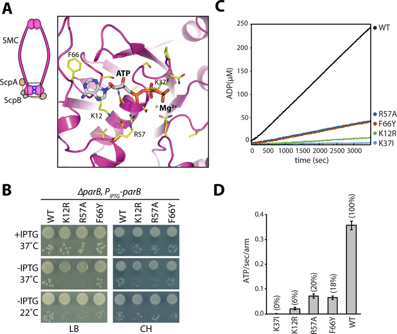
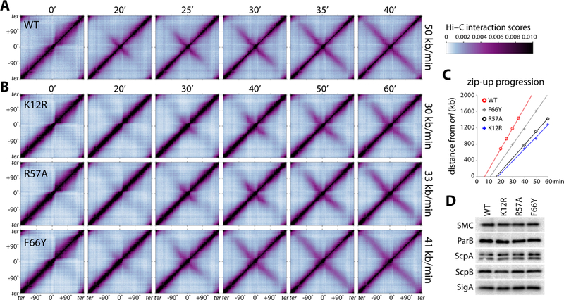
Structural maintenance of chromosomes (SMC) complexes shape the genomes of virtually all organisms, but how they function remains incompletely understood. Recent studies in bacteria and eukaryotes have led to a unifying model in which these ring-shaped ATPases act along contiguous DNA segments, processively enlarging DNA loops. In support of this model, single-molecule imaging experiments indicate that Saccharomyces cerevisiae condensin complexes can extrude DNA loops in an ATP-hydrolysis-dependent manner in vitro. Here, using time-resolved high-throughput chromosome conformation capture (Hi-C), we investigate the interplay between ATPase activity of the Bacillus subtilis SMC complex and loop formation in vivo. We show that point mutants in the SMC nucleotide-binding domain that impair but do not eliminate ATPase activity not only exhibit delays in de novo loop formation but also have reduced rates of processive loop enlargement. These data provide in vivo evidence that SMC complexes function as loop extruders.
Keywords: ParB; SMC; TAD; cohesin; condensin; loop extrusion.
Copyright © 2018 Elsevier Inc. All rights reserved.
Figures
Similar articles
-
Transient DNA Occupancy of the SMC Interarm Space in Prokaryotic Condensin.Mol Cell. 2019 Jul 25;75(2):209-223.e6. doi: 10.1016/j.molcel.2019.05.001. Epub 2019 Jun 11. Mol Cell. 2019. PMID: 31201090 Free PMC article.
-
Tuned SMC Arms Drive Chromosomal Loading of Prokaryotic Condensin.Mol Cell. 2017 Mar 2;65(5):861-872.e9. doi: 10.1016/j.molcel.2017.01.026. Epub 2017 Feb 23. Mol Cell. 2017. PMID: 28238653 Free PMC article.
-
Bacillus subtilis SMC complexes juxtapose chromosome arms as they travel from origin to terminus.Science. 2017 Feb 3;355(6324):524-527. doi: 10.1126/science.aai8982. Science. 2017. PMID: 28154080 Free PMC article.
-
Condensin complexes: understanding loop extrusion one conformational change at a time.Biochem Soc Trans. 2020 Oct 30;48(5):2089-2100. doi: 10.1042/BST20200241. Biochem Soc Trans. 2020. PMID: 33005926 Free PMC article. Review.
-
SMC complexes orchestrate the mitotic chromatin interaction landscape.Curr Genet. 2018 Apr;64(2):335-339. doi: 10.1007/s00294-017-0755-y. Epub 2017 Sep 21. Curr Genet. 2018. PMID: 28936767 Free PMC article. Review.
Cited by
-
Distinct Roles for Condensin's Two ATPase Sites in Chromosome Condensation.Mol Cell. 2019 Dec 5;76(5):724-737.e5. doi: 10.1016/j.molcel.2019.09.020. Epub 2019 Oct 16. Mol Cell. 2019. PMID: 31629658 Free PMC article.
-
The MukB-topoisomerase IV interaction mutually suppresses their catalytic activities.Nucleic Acids Res. 2022 Mar 21;50(5):2621-2634. doi: 10.1093/nar/gkab1027. Nucleic Acids Res. 2022. PMID: 34747485 Free PMC article.
-
Transient DNA Occupancy of the SMC Interarm Space in Prokaryotic Condensin.Mol Cell. 2019 Jul 25;75(2):209-223.e6. doi: 10.1016/j.molcel.2019.05.001. Epub 2019 Jun 11. Mol Cell. 2019. PMID: 31201090 Free PMC article.
-
Extrusion fountains are restricted by WAPL-dependent cohesin release and CTCF barriers.Nucleic Acids Res. 2025 Jun 20;53(12):gkaf549. doi: 10.1093/nar/gkaf549. Nucleic Acids Res. 2025. PMID: 40586309 Free PMC article.
-
High-Resolution 3D Genome Map of Brucella Chromosomes in Exponential and Stationary Phases.Microbiol Spectr. 2023 Feb 27;11(2):e0429022. doi: 10.1128/spectrum.04290-22. Online ahead of print. Microbiol Spectr. 2023. PMID: 36847551 Free PMC article.
References
-
- Burmann F, Shin HC, Basquin J, Soh YM, Gimenez-Oya V, Kim YG, Oh BH, and Gruber S (2013). An asymmetric SMC-kleisin bridge in prokaryotic condensin. Nat Struct Mol Biol 20, 371–379. - PubMed
Publication types
MeSH terms
Substances
Grants and funding
LinkOut - more resources
Full Text Sources
Other Literature Sources
Molecular Biology Databases
