. 2018 Sep 6;71(5):825-840.e6.
doi: 10.1016/j.molcel.2018.07.009. Epub 2018 Aug 9.

An Antiviral Branch of the IL-1 Signaling Pathway Restricts Immune-Evasive Virus Replication

Affiliations

An Antiviral Branch of the IL-1 Signaling Pathway Restricts Immune-Evasive Virus Replication

Megan H Orzalli et al. Mol Cell. .

Abstract

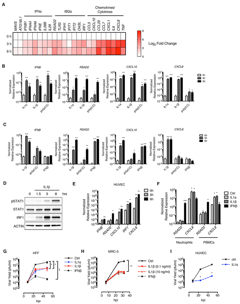
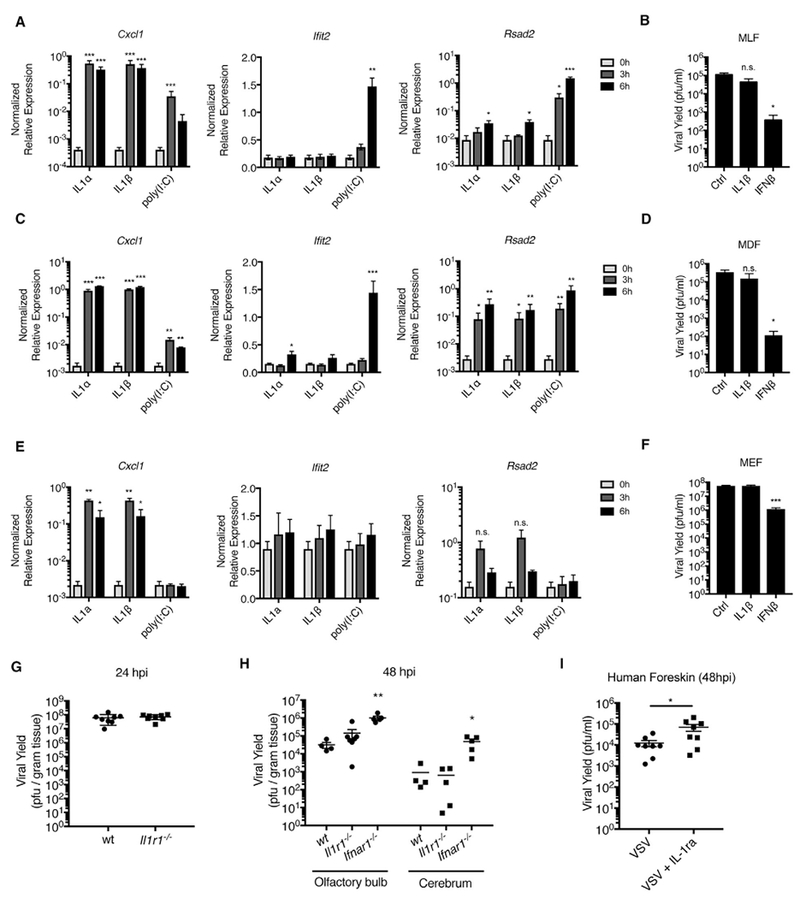
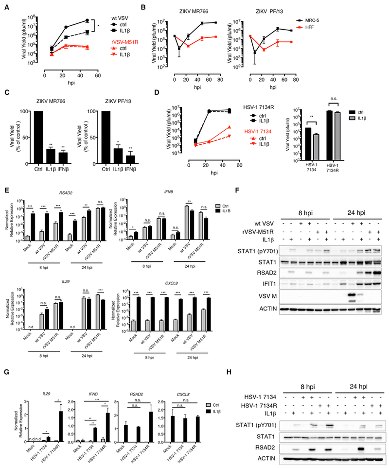
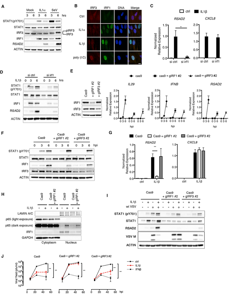
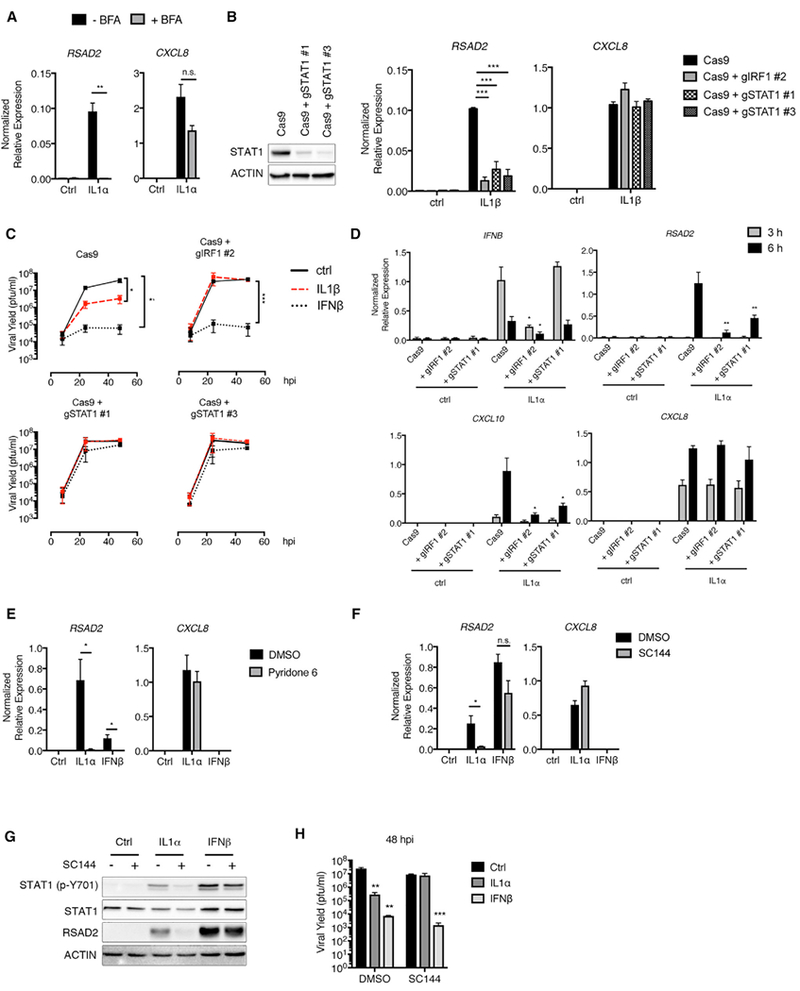
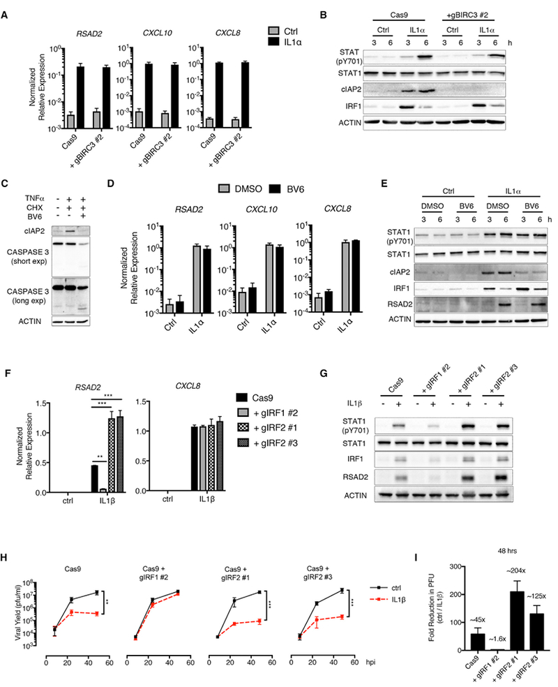
Virulent pathogens often cause the release of host-derived damage-associated molecular patterns (DAMPs) from infected cells. During encounters with immune-evasive viruses that block inflammatory gene expression, preformed DAMPs provide backup inflammatory signals that ensure protective immunity. Whether DAMPs exhibit additional backup defense activities is unknown. Herein, we report that viral infection of barrier epithelia (keratinocytes) elicits the release of preformed interleukin-1 (IL-1) family cytokines, including the DAMP IL-1α. Mechanistic studies revealed that IL-1 acts on skin fibroblasts to induce an interferon (IFN)-like state that restricts viral replication. We identified a branch in the IL-1 signaling pathway that induces IFN-stimulated gene expression in infected cells and found that IL-1 signaling is necessary to restrict viral replication in human skin explants. These activities are most important to control immune-evasive virus replication in fibroblasts and other barrier cell types. These findings highlight IL-1 as an important backup antiviral system to ensure barrier defense.

Keywords: IRF1; ISGs; antiviral defense; innate immunity; interferon regulatory factors.


Conflict of interest statement

DECLARATION OF INTERESTS

The authors declare no competing interests.

Figures

Figure 1.
Figure 1.. VSV Infection Elicits the Release of IL-1 Cytokines from Human Keratinocytes
(A–D) NOKs were mock-infected (ctrl) or infected with WT VSV or rVSV-M51R (MOI 10). (A) Total RNA was isolated at 8 hpi and analyzed by qRT-PCR. (B and D) Cell-free supernatants were harvested at indicated time points and analyzed by ELISA for IL-29 (B) and IL-1α/IL-1β (D). (C) Whole-cell lysates were harvested at indicated time points and separated by SDS-PAGE, and endogenous proteins were detected by immunoblot. (E) Total RNA was isolated from NOKs and HFFs. IL1A, IL1B, and IL1R1 transcript counts were quantified by nCounter and normalized to GAPDH. (F) 5 × 10⁵ NOKs or HFFs were lysed in 1% NP40 buffer and clarified by centrifugation, and ELISAs were used to analyze the soluble fractions for IL-1α and IL-1β. (G) NOKs and HFFs were stimulated with IL-1α or IL-1β (10 ng/mL). Total RNA was harvested at 6 hr and quantified by qRT-PCR. Samples are normalized to GAPDH and presented as fold induction over the control (ctrl) samples. (A–G) Data are the average of at least three independent experiments ± SEM. Student’s t test: *p < 0.05; **p < 0.01; ***p < 0.001.
Figure 2.
Figure 2.. IL-1 Cytokines Induce an Antiviral Response in Human Fibroblasts and Endothelial Cells
(A) HFFs were stimulated with recombinant IL-1β, and total RNA was isolated at 0, 3, and 6 hr post-stimulation. Absolute transcript counts were measured by nCounter and normalized to GAPDH. Data are presented as a fold induction over the 0 hr time point. (B and C) HFFs (B) and MRC-5 lung fibroblasts (C) were stimulated with IL-1α, IL-1β, poly I:C, or IFN β. Total RNA was isolated at indicated time points and analyzed by qRT-PCR. (D) Whole-cell lysates from IL-1β-stimulated HFFs were separated by SDS-PAGE and analyzed by immunoblot. (E) HUVECs were stimulated with IL-1α. Total RNA was isolated 0, 3, and 6 hr post-stimulation and analyzed by qRT-PCR. (F) Human neutrophils or peripheral blood mononuclear cells (PBMCs) were stimulated with IL-1α, IL-1β, or IFN β for 6 hr. Total RNA was isolated and analyzed by qRT-PCR. (G–I) HFFs (G), MRC-5 lung fibroblasts (H), and HUVECs (I) were infected with WT VSV (MOI 0.1) in the presence of IL-1α, IL-1β, or IFNβ. Cell-free supernatants were collected at 8, 24, 32, and/or 48 hpi, and viral yields were determined by plaque assay. (A–I) IL-1 cytokine stimulations were performed at 10 ng/mL unless otherwise stated. Poly I:C or IFN β stimulations were performed at 1 μg/mL or 10 U/mL, respectively. Data are the average of at least three independent experiments ± SEM. (E and I) Data are the average of two independent experiments ± SEM. Student’s t test: *p < 0.05; **p < 0.01; and ***p < 0.001.
Figure 3.
Figure 3.. IL-1 Does Not Restrict VSV Replication in Murine Fibroblasts
(A, C, and E) Mouse lung fibroblasts (MLFs) (A), mouse dermal fibroblasts (MDFs) (C), and mouse embryonic fibroblasts (MEFs) (E) were stimulated with IL-1α (10 ng/mL), IL-1β (10 ng/mL), or poly I:C (1 μg/mL). Total RNA was isolated at 0, 3, and 6 hr post-stimulation and analyzed by qRT-PCR. (B, D, and F) MLFs (B), MDFs(D), and MEFs (F) were infected with WT VSV (MOI 0.1) in the presence orabsenceofIL-1β (10ng/mL) or IFNβ (10U/mL). Viral yields were determined from infected-cell supernatants at 24 hpi by plaque assay. (G) Wild-type (wt) or Il1r1−/− mice were infected with 5 × 10⁶ plaque-forming units (PFUs) of VSV via epidermal scarification. Viral titer within the skin was determined at 24 hpi. (H) wt, Il1r1−/−, or Ifnar1−/− mice were infected with 5 × 10⁶ PFUs of VSV via intranasal injection. Viral titer was determined from indicated tissues at 48 hpi. (I) Punch biopsies (5 mm) from human newborn foreskin were infected with 2.5 × 10³ PFUs of VSV in the presence or absence of IL-1ra (500 ng) by intradermal injection. Results are the combined data points from two independent experiments ± SEM. (A–F) Results are an average of three independent experiments ± SEM. Student’s t test; *p < 0.05; **p < 0.01; and ***p < 0.01.
Figure 4.
Figure 4.. IL-1 Cytokines Restrict the Replication of Immune Evasive Viruses
(A) HFFs were infected with WT VSV or rVSV-M51R (MOI 0.1) in the presence of IL-1β. Cell-free supernatants were collected at 8, 24, and 48 hpi, and viral yields were determined by plaque assay. (B) HFF and MRC-5 fibroblasts were infected with ZIKV strains MR766 and PF/13 (MOI 1). Viral yields were determined as in (A) at 8, 24, 48, and 72 hpi. (C) MRC-5 lung fibroblasts were infected with indicated ZIKV strains in the presence ofIL-1β or IFNβ. Viral titers from infected-cell supernatants were determined at 48 hpi by plaque assay. Data are presented as a percentage of PFUs in control (ctrl)-treated cells. (D) HFFs were infected with HSV-1 7134 or 7134R (MOI 0.1). (Left panel) Virus was isolated from infected cells at 8, 24, and 48 hpi, and viral yields were determined by plaque assay on U2OS cells. (Right panel) Plaque assays were performed on infected-cell lysates isolated at 48 hpi. (E and F) HFFs were infected as described in (A). (E) Total RNA was isolated at 8 and 24 hpi and analyzed by qRT-PCR. Data are normalized to GAPDH. (F) Whole-cell lysates were isolated at 8 and 24 hpi, separated by SDS-PAGE, and analyzed by immunoblot. (G and H) HFFs were infected as described in (D). (G) Total RNA was isolated at 8 hpi and analyzed by qRT-PCR. Data are normalized to 18S rRNA. (H) Whole-cell lysates were isolated at 8 and 24 hpi, separated by SDS-PAGE, and analyzed by immunoblot. (A–H) IL-1 and IFNβ cytokine stimulations were performed at 10 ng/mL or 10 U/mL, respectively. Data are the average of three independent experiments ± SEM. (D, left panel) Data are representative of two independent experiments. Student’s t test: *p < 0.05; **p < 0.01; and ***p < 0.001.
Figure 5.
Figure 5.. IRF1 Is a Critical Regulator of IL-1-Mediated Antiviral Responses
(A) HFFs were stimulated with IL-1α (100 pg/mL) or infected with SeV (0.001 hemagglutinating units [HAUs]/cell). Whole-cell lysates were harvested at 3 and 6 hpi, separated by SDS-PAGE, and analyzed by immunoblot. (B) HFFs were stimulated with IL-1α (10ng/mL), IL-1β (10ng/mL), or poly(I:C) (1 μg/mL) for 6hr. Cells were fixed and stained with IRF3 and IRF1 antibodies. Nuclei were counterstained with DRAQ5. Data are representative of two independent experiments. The scale bar represents 10 μm. (C and D) HFFs were transfected with ctrl siRNAs or siRNAs targeting IRF1 for 72 hr. siRNA-transfected cells were then treated with IL-1β (10 ng/mL), and total RNA or protein were harvested at 3 and 6 hr or 6 hr, respectively. (C) Indicated transcripts were quantified by qRT-PCR and normalized to GAPDH. (D) Whole-cell lysates were separated by SDS-PAGE and analyzed by immunoblot. (E) (Left panel) Whole-cell lysates from HFFs transduced with lentiviruses expressing Cas9 alone or in combination with IRF1-or IRF3-targeted guide RNAs were separated by SDS-PAGE and analyzed by immunoblot. (Right panel) IRF1-and IRF3-deficient cells were infected SeV (0.001 HAU/cell). Total RNA was isolated from infected cells at 0, 3, and 6 hpi, and IL29, IFNB, and RSAD2 expressions were quantified by qRT-PCR. Samples were normalized to GAPDH. (F) Whole-cell lysates from SeV-infected Cas9 and IRF1- and IRF3-deficient cells were separated by SDS-PAGE and analyzed by immunoblot. (G) Cas9, IRF1-deficient, or IRF3-deficient HFFs were stimulated with IL-1β (10 ng/mL). Total RNA was harvested from cells at 6 hr and analyzed by qRT-PCR. (H) Cas9 or IRF1-deficient HFFs were stimulated with IL-1β (10 ng/mL). Cellular cytoplasmic and nuclear fractions were isolated at 1 hr post-stimulation and analyzed by immunoblot. (I and J) Indicated cell lines were infected with WT VSV (MOI 0.1) in the presence or absence of IL-1β (I and J) or IFNβ (J). (I) Whole-cell lysates were harvested at 24 hpi, separated by SDS-PAGE, and analyzed by immunoblot. (J) Infected-cell supernatants were harvested at indicated time points, and viral yields were determined by plaque assay. Data are the average of at least three independent experiments ± SEM. Student’s t test: *p < 0.05, **p < 0.01
Figure 6.
Figure 6.. Gp130 and STAT1 Are Essential for IL-1-Mediated Antiviral Responses
(A) HFFs were stimulated with IL-1α (100 pg/mL) in the presence or absence of brefeldin A (BFA) (1 μg/mL). Total RNA was isolated at 6 hr post-stimulation and analyzed by qRT-PCR. (B) (Left panel) STAT1 deficiency was demonstrated by the absence of STAT1 protein by immunoblot. (Right panel) Cells were stimulated with IL-1β (10 ng/mL).Total RNA or whole-cell lysates were harvested from cells at 6 hr and analyzed by qRT-PCR. (C) Cells were infected with WT VSV (MOI 0.1) in the presence or absence of IL-1β (10 ng/mL) or IFNβ (10 U/mL). Infected-cell supernatants were harvested at indicated time points, and viral yield was determined by plaque assay. (D) Cells were stimulated with IL-1α (100 pg/mL). Total RNA was harvested at 3 and 6 hr and analyzed by qRT-PCR. (E) HFFs pre-treated with DMSO or pyridone 6 (5 μg/mL) for 1 hr were stimulated with IL-1α (100 pg/mL) or IFNβ (1 U/mL). Total RNA was isolated at 6 hr and analyzed by qRT-PCR. (F and G) HFFs were pre-treated with a gp130 inhibitor (SC144; 25 μM) for 1 hr, prior to dilution with an equal volume of media containing IL-1α (200 pg/mL) or IFNβ (20 U/mL). Total RNA or whole-cell lysates were harvested at 6 hr and analyzed by (F) qRT-PCR or (G) separated by SDS-PAGE and analyzed by immunoblot. (H) SC144 (12.5 μM) alone or in combination with IL-1α (100 pg/mL) or IFNβ (10 U/mL) was added to cells 1 hr after infection with WT VSV (MOI 0.1). Infected-cell supernatants were isolated at 48 hpi and analyzed by plaque assay. Data are the average of at least three independent experiments ± SEM. Student’s t test: *p < 0.05; **p < 0.01; and ***p < 0.001.
Figure 7.
Figure 7.. IRF2 Negatively Regulates IL-1-Mediated Antiviral Responses
(A and B) Cas9 control or BIRC3-deficient fibroblasts were stimulated with IL-1α (100 pg/mL). (A) Total RNA was isolated at 6 hr and analyzed by qRT-PCR. (B) Whole-cell lysates were isolated at 3 and 6 hr post-stimulation, separated by SDS-PAGE, and analyzed by immunoblot. (C) HFFs were treated with cycloheximide (CHX) (10 μg/mL) and BV6 (5 μM) for 1 hr, followed by stimulation with TNF (10 ng/mL). Whole-cell lysates were isolated at 6 hr post-stimulation, separated by SDS-PAGE, and analyzed by immunoblot. (D and E) HFFs were pre-treated with BV6 as described in (C) and stimulated with IL-1α (100 pg/mL). (D) Total RNA was isolated at 6 hr post-stimulation and analyzed by qRT-PCR. (E) Whole-cell lysates were isolated at 3 and 6 hr post-stimulation, separated by SDS-PAGE, and analyzed by immunoblot. (F–I) Cas9 and IRF1- and IRF2-deficient HFFs were stimulated with (F and G) IL-1β (10 ng/mL) alone or (H–I) infected with WT VSV (MOI 0.1) in the presence or absence of IL-1β (10 ng/mL). (F) Total RNA was isolated at 6 hr post-stimulation and analyzed by qRT-PCR. (G) Whole-cell lysates were isolated at 6 hr, separated by SDS-PAGE, and analyzed by immunoblot. (H) Infected-cell supernatants were harvested at 8, 24, and 48 hpi, and viral yields were quantified by plaque assay. (I) The fold reduction in viral PFUs in IL-1b-treated cells (H) was determined at 48 hpi. Data are the average of at least three independent experiments ± SEM. Student’s t test: **p < 0.01; and ***p < 0.001.

References

    1. Ahmed M, McKenzie MO, Puckett S, Hojnacki M, Poliquin L, and Lyles DS (2003). Ability of the matrix protein of vesicular stomatitis virus to suppress beta interferon gene expression is genetically correlated with the inhibition of host RNA and protein synthesis. J. Virol 77, 4646–4657. - PMC - PubMed
    1. Alexopoulou L., Holt AC., Medzhitov R., and Flavell RA. (2001). Recognition of double-stranded RNA and activation of NF-kappaB by Toll-like receptor 3. Nature 413, 732–738. - PubMed
    1. Arpaia N, and Barton GM (2011). Toll-like receptors: key players in antiviral immunity. Curr. Opin. Virol 1, 447–454. - PMC - PubMed
    1. Auron PE, Webb AC, Rosenwasser LJ., Mucci SF, Rich A, Wolff SM, and Dinarello CA (1984). Nucleotide sequence of human monocyte interleukin 1 precursor cDNA. Proc. Natl. Acad. Sci. USA 81, 7907–7911. - PMC - PubMed
    1. Brien JD., Lazear HM., and Diamond MS. (2013). Propagation, quantification, detection, and storage of West Nile virus. Curr. Protoc. Microbiol 31, 15D.3.1.–15D.3.18. - PubMed

Publication types