. 2018 Aug;14(2):190-196.
doi: 10.5152/iao.2018.4974.

Atraumatic Scala Tympani Cochleostomy; Resolution of the Dilemma

Affiliations

Atraumatic Scala Tympani Cochleostomy; Resolution of the Dilemma

Ahmad Badr et al. J Int Adv Otol. 2018 Aug.

Abstract

Objectives: While an accurate placement in cochleostomy is critical to ensure appropriate insertion of the cochlear implant (CI) electrode into the scala tympani (ST), the choice of preferred cochleostomy sites widely varied among experienced surgeons. We present a novel technique for precise yet readily applicable localization of the optimum site for performing ST cochleostomy.

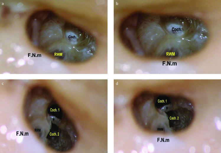
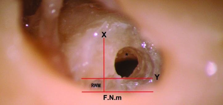
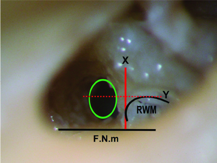
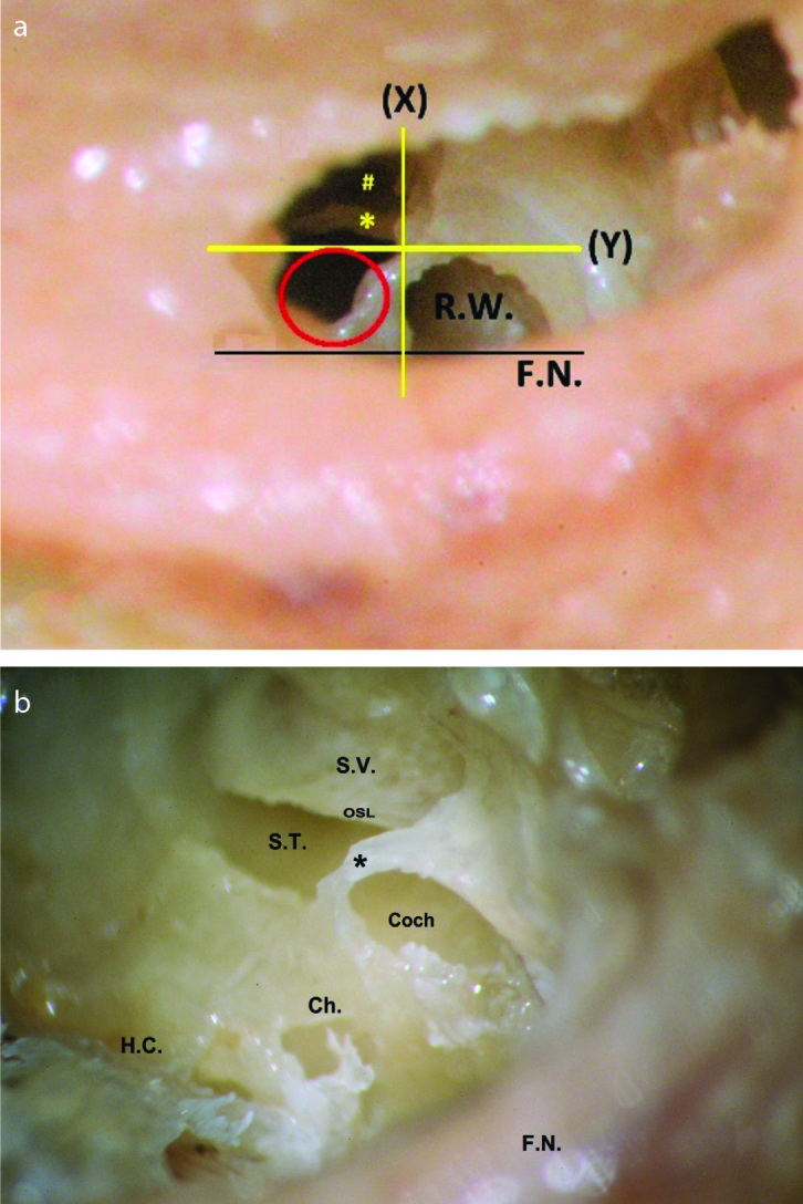
Material and methods: Twenty fresh frozen temporal bones were dissected using the mastoidectomy-posterior tympanotomy approach. Based on the facial nerve and the margins of the round window membrane (RWM), the cochleostomy site was chosen to insert the electrode into the ST while preserving the surrounding intracochlear structures.

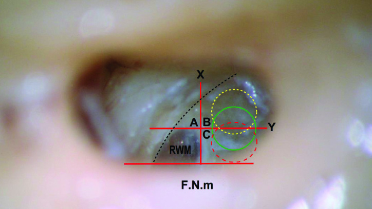
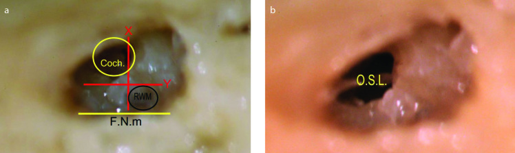
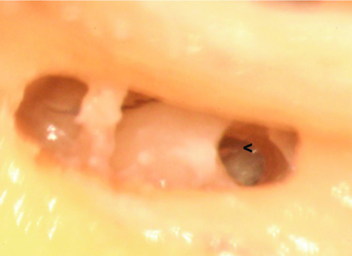
Results: There is a limited safe area suitable for the ST implantation in the area inferior and anterior to the RWM. There is a higher risk of scala vestibuli (SV) insertion anterior to that area. Posterior to that area, the cochlear aqueduct (CA) and inferior cochlear vein (ICV) are liable for the injury.

Conclusion: For atraumatic CI, precise and easy localization of the site of cochleostomy play a pivotal role in preserving intracochlear structures. Accurate setting of the vertical and horizontal orientations is mandatory before choosing the site of cochleostomy. The facial nerve and the margins of the RWM offer a very helpful clue for such localization; meanwhile, it is readily identifiable in the surgical field.


Conflict of interest statement

Conflict of Interest: The authors have no conflict of interest to declare.

Figures

Figure 1. a–d
Figure 1. a–d
The importance of proper orientation of the F.N.m. as seen during performing a right cochleostomy. This figure illustrates the importance of proper orientation of the mastoid segment of the facial nerve (F.N.m). It should lie exactly transversely across the surgical field, for accurate interpretation of the site of cochleostomy. (a) When the nerve was oblique across the field, misinterpretation of the site of the cochleostomy (Coch.) occurred, giving the impression of being anteroinferior to the round window membrane (RWM). Notice the preservation of the endosteal layer, to be later opened with a micro pick, not directly by the drill. (b) After proper transverse positioning of the nerve across the field, the site that seemed to be anteroinferior appears clearly now to be rather anterior to the RWM. N.B.: The rotation is obtained by editing the photo. (c) The endosteum was removed to discover that this cochleostomy site (Coch. 1) had led to the scala vestibuli supero-lateral to the osseous spiral lamina and basilar membrane (*). Then, a second cochleostomy (Coch. 2) was performed in an attempt to gain access to the scala tympani. During drilling, the cochleostomy seemed to be inferior to the RWM. (d) However, rotating the photo to have the facial nerve across the field demonstrated that the second cochleostomy lies anteroinferior to the RWM (rather than inferior).
Figure 2
Figure 2
The intermediate and safe-range cochleostomy. When the annulus of the right round window membrane (RWM) is exposed, imaginary tangents are considered touching the anterior and inferior parts of the annulus. Y line: the anterior tangent that is parallel to the mastoid segment of the facial nerve (F.N.m). X line: the inferior tangent that is vertical to the F.N.m. The X and Y lines divide the area anterior and inferior to the RWM into three areas: A, B, and C. Area A is the area anterior to the RWM, anterior to the Y line, and superior to the X line. Anterior cochleostomy shall lie in area A. Area B is the area anteroinferior to the RWM, inferior to the X line, and anterior to the Y line. Without the presence of a precise definition, cochleostomy in any part of area B can be designated as an anteroinferior cochleostomy. Area C is the area inferior to the RWM, inferior to the X line, and posterior to the Y line. Inferior cochleostomy shall be performed in area C. The green circle that is centered on the Y line and inferior to the X line marks the site of our recommended intermediate cochleostomy position. The term intermediate describes its interposition between the areas of the famous anteroinferior and inferior cochleostomies. The yellow fine-dashed circle represents the most anterior limit of the safe cochleostomy range, through which atraumatic scala tympani implantation can be performed, whereas the red coarse-dashed one represents the most posterior limit of that range. The dashed parabola represents the estimated course of the spiral ligament and osseous spiral lamina. Therefore, the area anterosuperior to this dashed parabola corresponds to the scala vestibuli, and the area postero-inferior to it corresponds to the scala tympani.
Figure 3. a, b
Figure 3. a, b
Left (traumatic) anteroinferior cochleostomy, leading to the scala tympani and the scala vestibuli. (a) Left anteroinferior cochleostomy. F.N.m: Facial nerve, mastoid segment. Coch.: Cochleostomy. (b) Cochleostomy led to the area of junction between the scala tympani and scala vestibuli. OSL: osseous spiral lamina
Figure 4
Figure 4
Right (traumatic) anteroinferior cochleostomy, leading mainly to the scala tympani and partially to the scala vestibuli. The osseous spiral lamina (<) is seen in the anterior one-fourth of the cochleostomy. Notice that the narrow space between the facial and chorda tympani nerves, together with the posterior rotation of the cochlea, prevented simultaneous visualization of the round window and the cochleostomy.
Figure 5
Figure 5
Right anteroinferior cochleostomy, touching the Y line and leading to the scala tympani immediately under the osseous spiral lamina (*). F.N.m: facial nerve, mastoid segment. RWM: anteroinferior part of the true round window membrane; the rest of the membrane is hidden medial to the facial nerve.
Figure 6
Figure 6
Left intermediate cochleostomy, the green circle is centered on the Y line and inferior to the X line. The cochleostomy purely led to the scala tympani; the osseous spiral lamina is not seen through the lumen of cochleostomy. F.N.m: facial nerve, mastoid segment; RWM: round window membrane
Figure 7. a, b
Figure 7. a, b
Frank inferior cochleostomy. (a) Posterior enlargement of the initial “intermediate cochleostomy” that was centered on the Y line, in an attempt to expose the scala tympani (ST) postero-inferior to the osseous spiral lamina (OSL) (*). (#) denotes the defect anterior to the OSL, after drilling the initial cochleostomy, which resulted in scala vestibuli (SV) insertion. Notice the position of the red circle (marking the ideal cochleostomy site, frank inferior cochleostomy in this case), in relation to the Y line, touching the line posteriorly. In addition, notice the steep vertical orientation of the OSL. RW: round window, the membrane was removed. F.N.: facial nerve. (b) The same specimen after lateral temporal bone resection and cochlear drill-out viewed from posterosuperior-lateral view. The basal cochlear turn has been opened to show the OSL, ST, and SV, with preservation of a bony rim (*) around the Coch. The channels for the cochlear aqueduct and the inferior cochlear vein (Ch.) seem very close to the posterior margin of the posterior extension. FN: facial nerve; HC: hypotympanic cells

References

    1. Alice B, Silvia M, Laura G, Patrizia T, Roberto B. Cochlear implantation in the elderly: surgical and hearing outcomes. BMC Surg. 2013;13(Suppl 2):S1. doi: 10.1186/1471-2482-13-S2-S1. - DOI - PMC - PubMed
    1. Marsot-Dupuch K, Meyer B. Cochlear implant assessment: imaging issues. Eur J Radiol. 2001;40:119–32. doi: 10.1016/S0720-048X(01)00380-1. - DOI - PubMed
    1. Campbell A, Dillon M, Buchman C, Adunka O. Hearing preservation cochlear implantation. Curr Otorhinolaryngol Rep. 2013;1:69–79. doi: 10.1007/s40136-013-0012-y. - DOI
    1. Kim LS, Jeong SW, Lee YM, Kim JS. Cochlear implantation in children. Auris Nasus Larynx. 2010;37:6–17. doi: 10.1016/j.anl.2009.09.011. - DOI - PubMed
    1. Lehnhardt E. Intracochlear placement of cochlear implant electrodes in soft surgery technique. HNO. 1993;7:356–9. - PubMed

MeSH terms