. 2018 Aug;14(2):312-316.
doi: 10.5152/iao.2018.5473.

Tricellulin Expression and its Deletion Effects in the Endolymphatic Sac

Affiliations

Tricellulin Expression and its Deletion Effects in the Endolymphatic Sac

Ai Matsubara et al. J Int Adv Otol. 2018 Aug.

Abstract

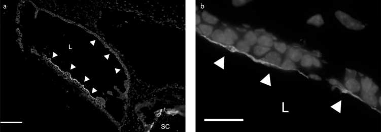
Objectives: Tricellulin is a tight junction (TJ)-forming protein that participates in the sealing function of tricellular TJs. Tricellulin-knockout (Tric-/-) mice show progressive hearing loss with degeneration of hair cells in the cochlea without physiological or physical disorders. In the present study, we investigated the tricellulin expression and its deletion effects in the endolymphatic sac (ES) using Tric-/- mice.

Materials and methods: The ES epithelia from wild-type (WT) mice were laser-microdissected, and RT-PCR was performed. The ES sections from Tric-/- and WT mice were immunostained with an anti-tricellulin antibody. Hematoxylin and eosin staining was performed for morphological examination. The inner ear of Tric-/- mice was perfused with biotinylation reagents, and the ES sections were observed for tracer permeability assay after applying streptavidin-Alexa Fluor 488 conjugate.

Results: The tricellulin expression was confirmed by RT-PCR and by immunohistochemistry in the WT ES. The ES in Tric-/- mice showed normal morphology and revealed no biotin leakage from the lumen.

Conclusion: The ES in Tric-/- mice showed no changes in morphology or disruption in macromolecular barrier function. The effects of solute leakages in the ES of Tric-/- mice may be very limited and compensatable, or that the ES epithelia may have other sealing system covering the lack of tricellulin.


Conflict of interest statement

Conflict of Interest: The authors have no conflict of interest to declare.

Figures

Figure 1
Figure 1
Agarose gel electrophoresis of PCR-amplified products from WT mice. A band for Tric was detected at the expected size in RNA isolated from the WT ES epithelia. The WT kidney template was used as a positive control. ES: endolymphatic sac; Tric: tricellulin; GAPDH: glyceraldehyde-3-phosphate dehydrogenase; M: measure; WT: wild-type.
Figure 2. a–f
Figure 2. a–f
Immunohistochemistry in the ES of WT (a–c) and Tric−/− mice (d–f). a, d. ZO-1 stained; b, e. tricellulin stained; c, f. merged images. In the ES epithelium from the WT mouse, tricellulin was stained at the corners of three-cell contacts (arrows in b and c). Tricellulin was not detected in the ES epithelium from the Tric−/− mouse (e). ZO-1 was double-stained red, showing linear expression around the cells (a and d). ES: endolymphatic sac; WT: wild-type; Tric−/−: tricellulin-knockout; ZO-1: zonula occludens-1. Scale bar: 20 μm.
Figure 3. a–c
Figure 3. a–c
Hematoxylin and eosin staining in the ES and cochlea of Tric−/− mice. a. ES in Tric−/− mice; b. ES in WT mice; c. cochlea in Tric−/− mice. The ES in Tric−/− mice (a) had no anatomical and morphological anomalies compared with the ES in WT mice (b). The epithelium was maintained in the regular one-layer sac structure. The intermediate portion of the ES was partially surrounded by bone (OP) and showed no apparent ballooning of the sac. The cochlea in Tric−/− mice had no findings of endolymphatic hydrops (c). ES: endolymphatic sac; Tric−/−: tricellulin-knockout; WT: wild-type; L: lumen of the ES; SC: semicircular canal; SV: scala vestibule; SM: scala media; ST: scala tympani; RM: Reissner’s membrane; OP: operculum. Scale bar: 100 μm.
Figure 4. a, b
Figure 4. a, b
Tracer permeability assay in Tric−/− mice. Biotin chemical compounds were perfused into the endolymph through the stria vascularis to the cochlear ducts. In the ES of Tric−/− mice, biotin was confined in the endolymph (arrowheads), and there was no leakage through the epithelia. Biotin was also detected in the lumen of the semicircular canal (a). Biotin was detected using a streptavidin–Alexa Fluor 488 conjugate. Tric−/−: tricellulin-knockout; ES: endolymphatic sac; L: lumen of the ES; SC: semicircular canal. Scale bar: a: 100 μm, b: 20 μm.

Similar articles

References

    1. Inamoto R, Miyashita T, Akiyama K, Mori T, Mori N. Endolymphatic sac is involved in the regulation of hydrostatic pressure of cochlear endolymph. Am J Physiol Regul Integr Comp Physiol. 2009;297:R1610–4. doi: 10.1152/ajpregu.00073.2009. - DOI - PubMed
    1. Salt AN, Plontke SK. Endolymphatic hydrops: pathophysiology and experimental models. Otolaryngol Clin North Am. 2010;43:971–83. doi: 10.1016/j.otc.2010.05.007. - DOI - PMC - PubMed
    1. Mori N, Miyashita T, Inamoto R, Matsubara A, Mori T, Akiyama K, et al. Ion transport its regulation in the endolymphatic sac: suggestions for clinical aspects of Meniere’s disease. Eur Arch Otorhinolaryngol. 2017;274:1813–20. doi: 10.1007/s00405-016-4362-1. - DOI - PMC - PubMed
    1. Staehelin LA. Structure and function of intercellular junctions. Int Rev Cytol. 1974;39:191–283. doi: 10.1016/S0074-7696(08)60940-7. - DOI - PubMed
    1. Bagger-Sjoback D, Rask-Andersen H. The permeability barrier of the endolymphatic sac. A hypothesis of fluid and electrolyte exchange based on freeze fracturing. Am J Otol. 1986;7:134–40. - PubMed

MeSH terms

Substances

LinkOut - more resources