. 2018 Aug 13;7(8):59.
doi: 10.1038/s41389-018-0069-z.

Notch3 inhibits epithelial-mesenchymal transition in breast cancer via a novel mechanism, upregulation of GATA-3 expression

Affiliations

Notch3 inhibits epithelial-mesenchymal transition in breast cancer via a novel mechanism, upregulation of GATA-3 expression

Hao-Yu Lin et al. Oncogenesis. .

Abstract

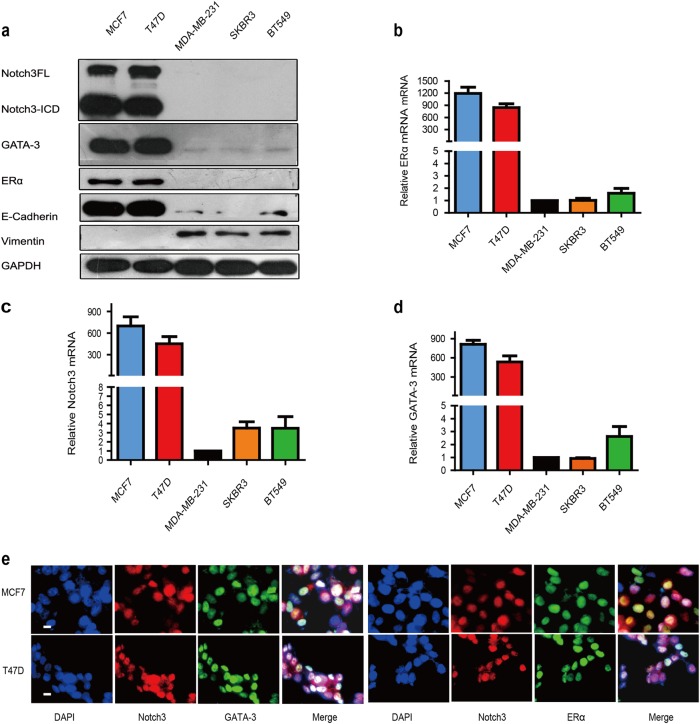
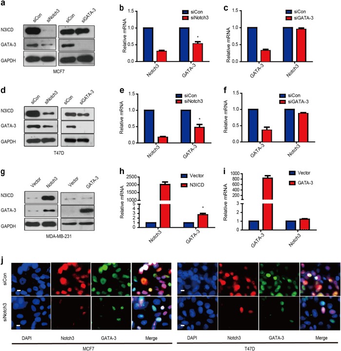
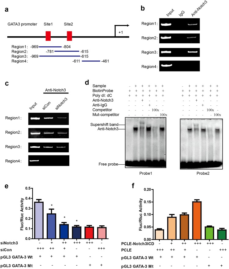
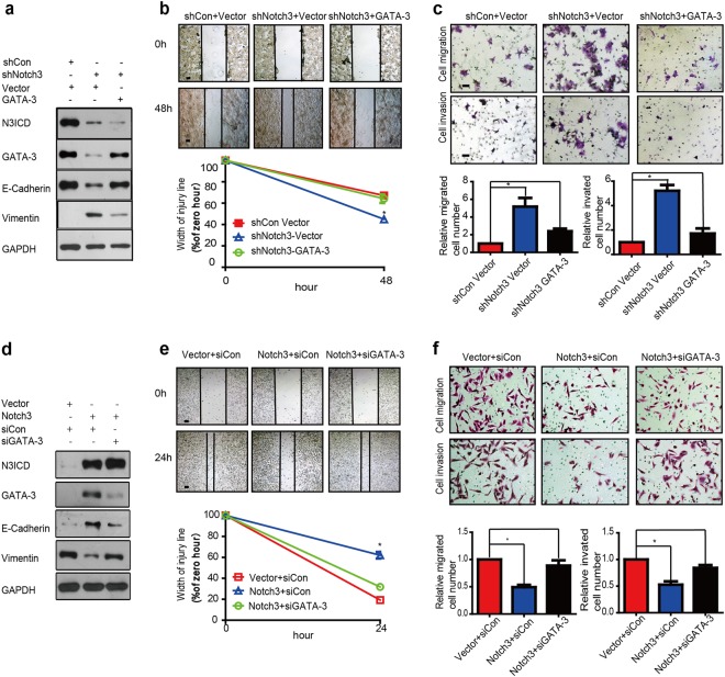
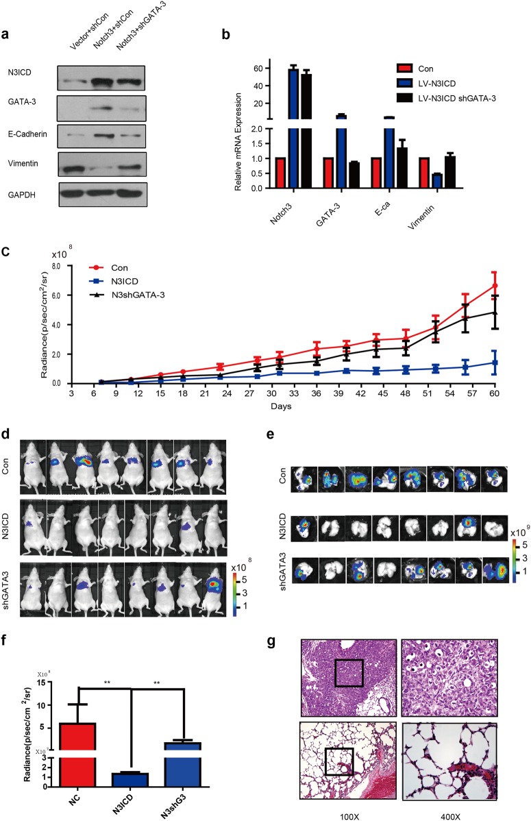
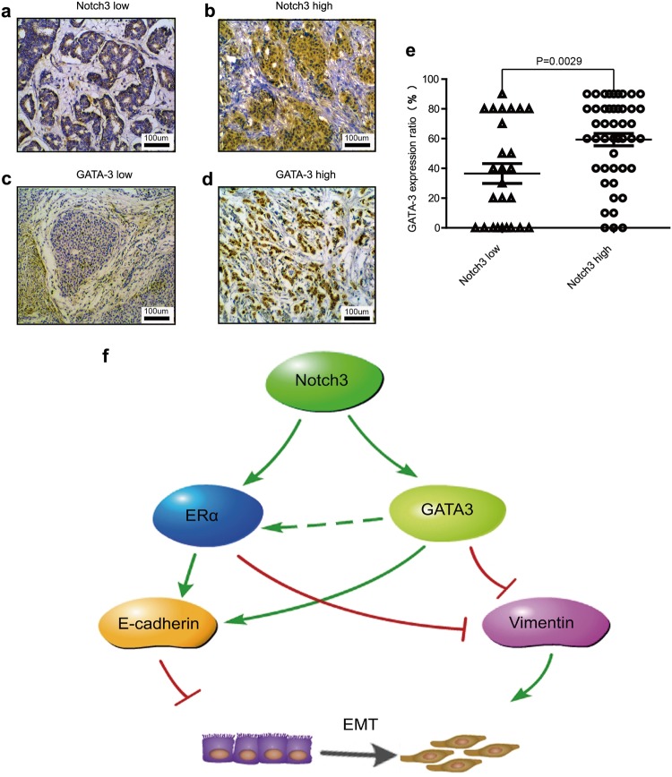
Notch3 and GATA binding protein 3 (GATA-3) have been, individually, shown to maintain luminal phenotype and inhibit epithelial-mesenchymal transition (EMT) in breast cancers. In the present study, we report that Notch3 expression positively correlates with that of GATA-3, and both are associated with estrogen receptor-α (ERα) expression in breast cancer cells. We demonstrate in vitro and in vivo that Notch3 suppressed EMT and breast cancer metastasis by activating GATA-3 transcription. Furthermore, Notch3 knockdown downregulated GATA-3 and promoted EMT; while overexpression of Notch3 intracellular domain upregulated GATA-3 and inhibited EMT, leading to a suppression of metastasis in vivo. Moreover, inhibition or overexpression of GATA-3 partially reversed EMT or mesenchymal-epithelial transition induced by Notch3 alterations. In breast cancer patients, high GATA-3 expression is associated with higher Notch3 expression and lower lymph node metastasis, especially for hormone receptor (HR) positive cancers. Herein, we demonstrate a novel mechanism whereby Notch3 inhibit EMT by transcriptionally upregulating GATA-3 expression, at least in part, leading to the suppression of cancer metastasis in breast cancers. Our findings expand our current knowledge on Notch3 and GATA-3's roles in breast cancer metastasis.


Conflict of interest statement

The authors declare that they have no conflict of interest.

Figures

Fig. 1
Fig. 1. Correlation of Notch3 and GATA-3 expression in various breast cancer cell lines.
a Expression of Notch3, GATA-3, ERα, E-cadherin, and vimentin analyzed by western blot in breast cancer cell lines. bd Expression of Notch3, GATA-3, and ERα mRNA analyzed by reverse transcription (RT)–PCR in breast cancer cell lines. e Confocal fluorescence microscopy of DAPI/Notch3/GATA-3 and DAPI/Notch3/ERα staining in MCF-7 (top) and T47D (bottom) cells. The scale bar represents 200 μm
Fig. 2
Fig. 2. Notch3 regulates expression of GATA-3 transcriptionally.
ac Expression of Notch3 and GATA-3 in MCF-7 cells at protein and mRNA levels analyzed by western blot (a) and RT–PCR, respectively, bc when silencing Notch3 or GATA-3 by siRNA (df). Expression of Notch3 and GATA-3 in T47D cells at protein and mRNA levels analyzed by western blot (d) and RT–PCR (ef), respectively, when silencing Notch3 or GATA-3 by siRNA. gi Expression of Notch3 and GATA-3 in MDA-MB-231 cells at protein and mRNA levels analyzed by western blot (g) and RT–PCR, respectively, (hi) when overexpressing N3ICD or GATA-3. j Confocal fluorescence microscopy of DAPI/Notch3/GATA-3 staining in MCF-7 (left) and T47D (right) cells, treated with siControl (top) or siNotch3 (bottom). The scale bar represents 200 μm
Fig. 3
Fig. 3. Notch3 upregulates GATA-3 expression by binding to the core element of the GATA-3 promoter.
ac Schematic representation of the three CSL-binding element-containing primers (Regions 1 and 2 containing respective single CSL-binding elements, region 3 containing both CSL-binding elements) and negative control primers (region 1 does not contain a CSL-binding element) used for ChIP assays. Bands were seen in regions 1, 2, and 3 of the PCR product but not in the negative control region 4 (b); bands were dramatically decreased in regions 1, 2, and 3 of the PCR product when cells were treated with siNotch3 (c). d Probes 1 and 2 represent two biotin probes containing the core element of the CSL-binding sites of the GATA-3 promoter. Supershift bands (lane 3) were seen in the presence of an anti-Notch3 antibody, but not in the presence of an anti-IgG antibody (lane 4). Competition assays were performed using a 100-fold (lane 5) excess of unlabeled oligonucleotide containing the core element of the CSL-binding element of the GATA-3 promoter. A 100-fold excess of unlabeled mutation oligonucleotide containing the mutated core element of the CSL-binding element (lane 6) was used for mutation competition assays. Nuclear extracts from MCF-7 cells were not added to lane 1. e Notch3 was silenced in MCF-7 cells by siRNA and then co-transfected with the GATA-3 promoter or a mutated GATA-3 promoter (deleting both CLS-binding elements) construct containing Firefly luciferase, and internal control plasmid, pRL-SV40, containing Renilla luciferase. Firefly luciferase/Renilla luciferase values were used to indicate promoter activity. Each sample was tested in triplicate. *P < 0.05. f N3ICD was overexpressed in MDA-MB-231 cells and co-transfected with the GATA-3 promoter or a mutated GATA-3 promoter (deleting both CSL-binding elements) construct containing Firefly luciferase and internal control plasmid, pRL-SV40, containing Renilla luciferase. Firefly luciferase/Renilla luciferase values were used to indicate promoter activity. Each sample was tested in triplicate. *P < 0.05
Fig. 4
Fig. 4. Notch3 knockdown in MCF-7 cells promotes EMT while N3ICD overexpression in MDA-MB-231 cells inhibits EMT.
a Expression of Notch3, GATA-3, E-cadherin, and vimentin in MCF-7 cells analyzed by western blot when stably silencing Notch3, with or without overexpression of GATA-3. b Wound healing assay showed the knockdown of Notch3 enhanced the cellular motility of MCF-7 cells; such changes could be rescued by simultaneously adding a GATA-3 plasmid. Representative pictures (top) and quantitative data (bottom) of wound recovery after 48 h cell culture. Each sample contained three wells. c Representative pictures (top) and quantitative data (bottom) of migration or invasion assays. Each sample contained three wells. d Expression of Notch3, GATA-3, E-cadherin, and vimentin in MDA-MB-231 cells analyzed by western blot when stably overexpressing N3ICD, with or without knockdown of GATA-3 by siRNA. e Representative pictures (top) and quantitative data (bottom) of wound recovery after 24 h cell culture. Each sample contained three wells. f Representative pictures (top) and quantitative data (bottom) of migration or invasion assays. Each sample contained three wells. All experiments were performed at least three times and data were statistically analyzed by two-sided t-test. *P < 0.05, **P < 0.01, ***P < 0.001 vs. control. Error bars indicate standard error of the mean (SEM)
Fig. 5
Fig. 5. Notch3 inhibits distant metastasis via regulation of GATA-3 in a humanized mouse model.
ab Expression of Notch3, GATA-3, E-cadherin, and vimentin in MDA-MB-231 cells analyzed by western blot (a) or RT–PCR (b) when stably overexpressing N3ICD, with or without knockdown of GATA-3 by lentivirus infection. cd NU/NU mice were injected intravenously with MDA-MB-231 cells (1 × 106 cells) for the control group, Notch3 stably expressing group, or the stably expressing Notch3 with stably knocked down GATA-3 group. Bioluminescence imaging was performed 1 h after injection and then twice a week (c). The photon intensities of lung metastases in vivo in the three groups are indicated (mean ± SEM) (d). ef Mice were sacrificed to examine metastases 2 months after injection. Representative bioluminescence imaging was performed and mice were euthanized immediately after imaging (n = 8 independent mice per group). Bioluminescence imaging of a mouse lung which was taken out within 5 min of sacrifice (e). The photon intensities of lungs in vitro in the three groups are indicated (f) (mean ± SEM). g Representative hematoxylin and eosin (HE) staining for lung metastases in the two groups (Magnification: ×100 and ×400
Fig. 6
Fig. 6. Notch3 expression is associated with GATA-3 in breast cancer clinical cases.
ad Representative pictures of Notch3 low expression (a), Notch3 high expression (b), GATA-3 low expression (c), and GATA-3 high expression (d). Samples were stained by immunohistochemistry. e Positive correlation between Notch3 and GATA-3 expression (P = 0.0029) analyzed by two-sided t-test. *P < 0.05, **P < 0.01, ***P < 0.001 vs. control. f Proposed model of how Notch3 and GATA-3 regulate EMT in breast cancer

Similar articles

Cited by

References

    1. Siegel RL, Miller KD, Jemal A. Cancer Statistics, 2017. CA Cancer J. Clin. 2017;67:7–30. doi: 10.3322/caac.21387. - DOI - PubMed
    1. Bresnick EH, Lee HY, Fujiwara T, Johnson KD, Keles S. GATA switches as developmental drivers. J. Biol. Chem. 2010;285:31087–31093. doi: 10.1074/jbc.R110.159079. - DOI - PMC - PubMed
    1. Fortini ME. Introduction—Notch in development and disease. Semin. Cell Dev. Biol. 2012;23:419–420. doi: 10.1016/j.semcdb.2012.03.001. - DOI - PubMed
    1. Zheng R, Blobel GA. GATA transcription factors and cancer. Genes Cancer. 2010;1:1178–1188. doi: 10.1177/1947601911404223. - DOI - PMC - PubMed
    1. Hoyler T, et al. The transcription factor GATA-3 controls cell fate and maintenance of type 2 innate lymphoid cells. Immunity. 2012;37:634–648. doi: 10.1016/j.immuni.2012.06.020. - DOI - PMC - PubMed