. 2018 May;21(4):362-376.
doi: 10.1016/j.mattod.2017.10.005. Epub 2017 Nov 4.

Engineering vascularized and innervated bone biomaterials for improved skeletal tissue regeneration

Affiliations

Engineering vascularized and innervated bone biomaterials for improved skeletal tissue regeneration

Alessandra Marrella et al. Mater Today (Kidlington). 2018 May.

Abstract

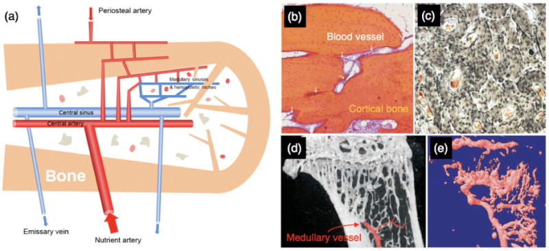
Blood vessels and nerve fibers are distributed throughout the entirety of skeletal tissue, and play important roles during bone development and fracture healing by supplying oxygen, nutrients, and cells. However, despite the successful development of bone mimetic materials that can replace damaged bone from a structural point of view, most of the available bone biomaterials often do not induce sufficient formation of blood vessels and nerves. In part, this is due to the difficulty of integrating and regulating multiple tissue types within artificial materials, which causes a gap between native skeletal tissue. Therefore, understanding the anatomy and underlying interaction mechanisms of blood vessels and nerve fibers in skeletal tissue is important to develop biomaterials that can recapitulate its complex microenvironment. In this perspective, we highlight the structure and osteogenic functions of the vascular and nervous system in bone, in a coupled manner. In addition, we discuss important design criteria for engineering vascularized, innervated, and neurovascularized bone implant materials, as well as recent advances in the development of such biomaterials. We expect that bone implant materials with neurovascularized networks can more accurately mimic native skeletal tissue and improve the regeneration of bone tissue.

Keywords: Bone biomaterials; innervated bone materials; neurovascularized bone materials; vascularized bone materials.


Figures

Figure 1
Figure 1
The vascular system in bone. (a) Schematic diagram of hierarchically structured blood vessels in bone. Blood supply is derived from medullary arteries, which are connected to periosteal arterial blood vessels. Medullary blood then exists via veins that penetrate the bone cortex. (b) Light micrograph image showing blood vessels penetrating cortical human bone (image width: 1.5 mm). (c) Transverse sectional view of human bone showing blood vessel penetration at the center of Haversian canals. (d) MicroCT image of mouse tibia showing the medullary vessels using a radio-opaque contrast agent (image width: 3.5 mm). (e) 3D rendered image of the blood vessel network of the same mouse tibia from panel (d), after a decalcification process (Images reproduced with permission from [38]).
Figure 2
Figure 2
Developmental angiogenesis in murine femur bone. (a) Confocal images showing blood vessel formation during the early stages of bone development, based on endomucin (Emcn)-stained endothelial cells. E: embryonic day, P: postnatal day. mp: metaphysis, dp: diaphysis. (b) Osterix immunostaining images illustrating skeletal development (Image reproduced with permission from [49]).
Figure 3
Figure 3
Schematic illustration of material properties that influence osteogenic differentiation and vascularization. (a) Osteogenic cells (blue) and vasculogenic cells (red) residing in the vascularized bone niche. (b) Material stiffness influences cellular adhesion, shape, and differentiation behaviour, based on mechanosensing of cells. (c) Materials that have nanoscale roughness can promote the attachment and differentiation of osteogenic and vasculogenic cells. (d) Generating an optimal level of porosity in bone materials is important to induce blood vessel growth, and maintain high mechanical strength of biomaterials.
Figure 4
Figure 4
(a) An integrated tissue-organ printer used to develop microchannels for inducing bone vascularization. (b) A 3D architecture with basic patterning composed of multiple types of cell-laden hydrogels and PCL polymers. (c) (i) Motion program used for printing 3D scaffolds as a calvarial bone substitute. Green colors represent the PCL/TCP dispensing paths, while red colors indicate the cell-laden hydrogel paths. (ii) Photograph (bottom) and SEM image (top) of the printed calvarial bone scaffold. (iii) Images of the printed scaffold at day zero (top) and 5 months after implantation (bottom). (iv–vi) H&E staining representing bone formation of non-treated (iv), scaffolds without cells (v) and hAFSCs-printed constructs (vi) 5 months after implantation. (vii–Modified tetrachrome staining of non-treated (vii), scaffolds without cells (viii) and hAFSCs-printed constructs (ix): red represents mature bone, and blue represents osteoid and lining of lacunae. (x–xii) vWF immunostaining of non-treated (x), scaffolds without cells (xi) and hAFSCs-printed constructs (xii): red parts show blood vessels, NB indicates new bone, while PCL/TCP indicates remaining scaffold (Images reproduced with permission from [110]).
Figure 5
Figure 5
Nervous system in bone. (a) The human peripheral nervous network in bone is connected to the central nervous system (CNS). Peripheral nervous system (PNS) in bone tissue transmits signals from external stimuli to CNS through the spinal cord (Image reproduced with permission from [112]). (b) Schematic image showing distribution of sensory neurons that innervate bone with different receptor types of sensory neurons.
Figure 6
Figure 6
Material properties that influence growth and differentiation of osteogenic cells and neurogenic cells. (a) Schematic illustration of innervated bone niche. (b) Nerve scaffolds and bone scaffolds require different stiffness levels to mimic native tissues. (c) Neurogenic differentiation of MSCs occurred in soft gel scaffolds (< ~1kPa) as determined by the expression of ??3 tubulin neuronal cytoskeletal marker. On the other hand, osteogenic differentiation of MSCs was induced when cells were cultured in stiff gel scaffolds (> ~25 kPa) as determined by expression of CBFa1 osteoblast transcription factor (arrow). Scale bar: 5μm. (d) From the immunostaining images and Western blotting images, neuronal markers were only expressed when MSCs were cultured in soft gel materials (< ~1 kPa). (e) Secretion of osteogenic proteins by MSCs was enhanced when cells were cultured in relatively stiff gel materials (> ~25 kPa). (Images reproduced with the permission of [74, 75]). (f) Roughness change at the nanoscale can significantly decrease the adhesion level of neuroblastoma cells. Left: AFM images of gold substrate with flat surfaces (Ra=0.46 nm) and nanorough surfaces (Ra=99.8 nm). Ra is the mean surface roughness. Right: Neuroblastoma cells grown on flat surfaces had improved adhesion and spreading morphology compared to nanorough surfaces. (Image reproduced with the permission of [161]). (g) Porosity is another critical material property affecting the adhesion behaviour of neuronal cells. Left: AFM images of macroporous and mesoporous silicon substrates. Middle and right: From immunostaining images and SEM images, neuroblastoma cells grown on mesoporous substrates showed better spreading morphology than macroporous substrates. (Image reproduced with the permission of [166]).
Figure 7
Figure 7
Magnesium ions (Mg2+) can enhance CGRP neuronal production to promote bone regeneration. (a) Photo image of Mg rod implanted in a rat cortical bone, from a cross-sectional view. (b–c) H&E staining images (b) and calcein-green labeling images (c) showing increase of new bone formation after implantation of Mg rod for 2 weeks (BM: bone marrow, NB: new bone, OB: old bone, and P: periosteum. Scale bar: 200 μm). (d) CGRP immunofluorescence staining images in DRGs in rat lumbar, 2 weeks post Mg rod implantation. Cellular nuclei are stained with DAPI (Scale bar: 50 μm). (e) Radiograph images of the fractured rat femur bone, 4 weeks post implantation of intramedullary nail with/without Mg component. To confirm the effect of CGRP, Ramp1 was knocked down or overexpressed in vivo (IMN: innovative intramedullary nail, AdV-NC: a negative control treated with scrambled adenoviruses, AdV-Ramp1: a group with Ramp1 overexpression, and AdV-shRamp1: a group with knocked down of Ramp1). (f) Schematic diagram explaining the effect of (Mg2+) on coupled osteogenesis and neurogenesis. Implant derived Mg2+ ions can enter DRG neurons through Mg2+ channels or transporters, enhancing accumulation and exocytosis of CGRP-vesicles. The CGRPs released from DRGs then trigger the CGRP receptor on PDSCs and induce osteogenic differentiation (Images reproduced with permission from [20]).
Figure 8
Figure 8
(a) Vessels (red) and nerves (green) aligned together (reproduced with permission from ref. [24]). (b–c) Immunohistochemistry of nerve markers in metaphysis (b) and deep metaphysis (c) of neonatal rat femur bone. Positive nerve marker staining (arrows), local bone trabaeculae (t) and blood vessels (V), osteoclasts (Oc), osteoblasts (Ob) and hematopoietic cells (H) are shown. Original magnification: 1000× for panel (b); 1500× for panel (c). (Images reproduced with permission from [167]). (d) Role of β-NGF on angiogenesis. β-NGF binds the TRk-A receptor on endothelial cell surface and sensory nerves, inducing SP and CGRP-I release from sensory nerves, which target endothelial cells through the NK1 and CGRPR receptors. β-NGF also binds to macrophage surface to induce VEGF release. (Image reproduced with permission from [23]).

Similar articles

Cited by

References

    1. Marsell R, Einhorn TA. Injury. 2011;42:551. - PMC - PubMed
    1. Das A, et al. Biomaterials. 2013;34:9853. - PMC - PubMed
    1. Dimitriou R, et al. BMC Med. 2011;9:66. - PMC - PubMed
    1. Calori GM, et al. Injury. 2007;38:11–18. - PubMed
    1. Verrier S, et al. Eur Cells Mater. 2016 - PubMed

LinkOut - more resources