. 2018 Jul 27:9:1735.
doi: 10.3389/fimmu.2018.01735. eCollection 2018.

β-Defensin 1 Is Prominent in the Liver and Induced During Cholestasis by Bilirubin and Bile Acids via Farnesoid X Receptor and Constitutive Androstane Receptor

Affiliations

β-Defensin 1 Is Prominent in the Liver and Induced During Cholestasis by Bilirubin and Bile Acids via Farnesoid X Receptor and Constitutive Androstane Receptor

Thomas Klag et al. Front Immunol. .

Abstract

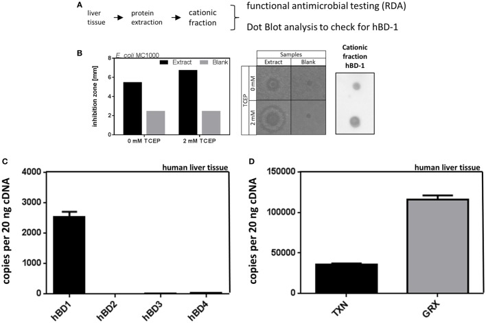
Background & aims: Knowledge about innate antimicrobial defense of the liver is limited. We investigated hepatic expression and regulation of antimicrobial peptides with focus on the human beta defensin-1 (hBD-1).

Methods: Radial diffusion assay was used to analyze antimicrobial activity of liver tissue. Different defensins including hBD-1 and its activator thioredoxin-1 (TXN) were analyzed in healthy and cholestatic liver samples by qPCR and immunostaining. Regulation of hBD-1 expression was studied in vitro and in vivo using bile duct-ligated mice. Regulation of hBD-1 via bilirubin and bile acids (BAs) was studied using siRNA.

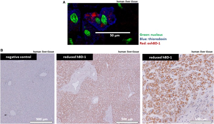
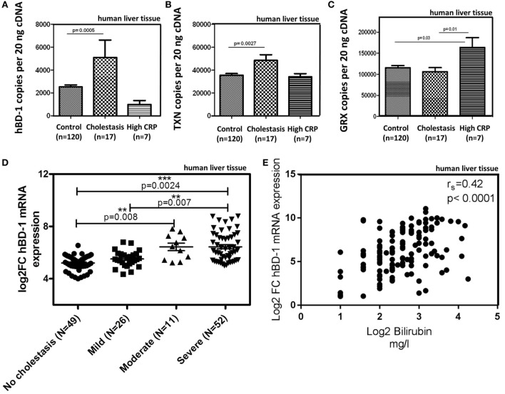
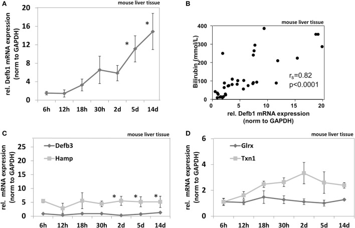
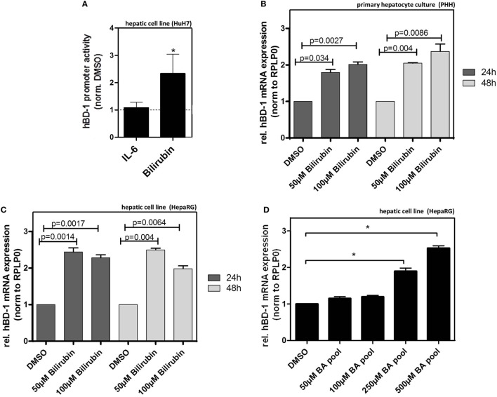
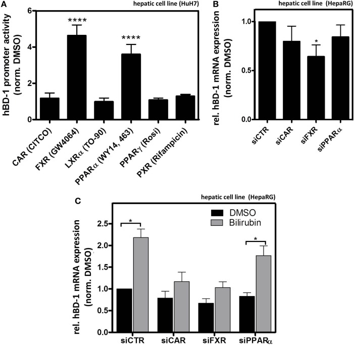
Results: We found strong antimicrobial activity of liver tissue against Escherichia coli. As a potential mediator of this antimicrobial activity we detected high expression of hBD-1 and TXN in hepatocytes, whereas other defensins were minimally expressed. Using a specific antibody for the reduced, antimicrobially active form of hBD-1 we found hBD-1 in co-localization with TXN within hepatocytes. hBD-1 was upregulated in cholestasis in a graded fashion. In cholestatic mice hepatic AMP expression (Defb-1 and Hamp) was enhanced. Bilirubin and BAs were able to induce hBD-1 in hepatic cell cultures in vitro. Treatment with siRNA and/or agonists demonstrated that the farnesoid X receptor (FXR) mediates basal expression of hBD-1, whereas both constitutive androstane receptor (CAR) and FXR seem to be responsible for the induction of hBD-1 by bilirubin.

Conclusion: hBD-1 is prominently expressed in hepatocytes. It is induced during cholestasis through bilirubin and BAs, mediated by CAR and especially FXR. Reduction by TXN activates hBD-1 to a potential key player in innate antimicrobial defense of the liver.

Keywords: antimicrobial peptides; bilirubin; cholestasis; hepatocytes; human β-defensin-1.


Figures

Figure 1
Figure 1
Antimicrobial activity of liver protein extract is accompanied by high expression of hBD-1 and oxidoreductases in human liver tissue. (A) Schematic workflow of cationic protein extraction from liver tissue, which was tested for antimicrobial activity [radial diffusion assay (RDA)] and hBD-1 status (by dot blot analysis). (B) On the left panel, quantitative results of RDA inhibition zones of cationic protein extract and blank extract (without liver tissue) under reducing (2 mM TCEP) or non-reducing conditions (0 mM TCEP) are depicted. In the middle panel, the respective RDA results are shown as picture. On the right panel, dot blot results with hBD-1 primary antibody to confirm hBD-1 peptide in cationic protein extract are shown. (C) mRNA expression of hBD-1, hBD-2, -3, and -4 was analyzed in a human patient cohort without cholestasis or systemic inflammatory response (n = 120). mRNA transcript levels are measured in an amount of 20 ng cDNA. Total copy numbers are depicted. Data are presented as means ± SEM for each gene assay. (D) mRNA expression of TXN and GRX in human liver tissue (n = 120). mRNA transcript levels are measured in an amount of 20 ng cDNA. Total copy numbers are depicted. Data are presented as means ± SEM.
Figure 2
Figure 2
Antimicrobial active—reduced hBD-1—is strongly expressed in human hepatocytes. (A) Liver tissue from a representative healthy person was stained for TRX (blue) and oxidized hBD-1 (red), showing hBD-1 and TXN being co-localized in human hepatocytes. Nuclear staining is depicted in green. (B) Staining of reduced hBD-1 shows a strong and ubiquitous expression of reduced hBD-1 in human liver tissue.
Figure 3
Figure 3
hBD-1 expression in samples of patients with cholestasis. (A–C) mRNA expression of hBD-1 (A) and human oxidoreductases TXN (B) and GRX (C) were analyzed in human liver tissue (n = 144). Control group (n = 120) without cholestasis was compared with cholestatic samples (serum bilirubin > 1.2 mg/dl, mean 4.5 mg/dl) and samples with signs of systemic inflammatory response (CRP > 5 mg/l). mRNA transcript levels are measured in an amount of 20 ng cDNA. Total copy numbers are depicted. Data are presented as means ± SEM. Values between groups were considered statistically significant with p < 0.05. (D) Hepatic hBD-1 mRNA expression was assessed in a cohort of patients with different grades of cholestasis (n = 138). Depicted are scatter blots of individual hBD-1 expression with horizontal lines indicating medians. Data were analyzed by Mann–Whitney test. **p < 0.01; ***p < 0.005. (E) Correlation matrix between mRNA expression of hBD-1 and bilirubin serum values. Spearman’s rank correlation coefficient is indicated as rs. Statistical significance for comparison was p < 0.0001.
Figure 4
Figure 4
In vivo expression of murine antimicrobial peptides in liver homogenates of bile duct-ligated mice. Gene expression pattern of Defb1 (A), Defb3 and Hamp (C), as well as Glrx and Txn1 (D) at multiple time points after bile duct ligation. Gene expression levels are shown as fold changes to sham-operated mice (0 h), normalized to GAPDH levels as average of 5 mice per time point. Error bars indicate SDs. Data were analyzed by student’s t-test; *p < 0.01. (B) Correlation matrix between mRNA expression of Defb1 and bilirubin values. Spearman’s rank correlation coefficient is indicated as rs. Statistical significance for comparison was p < 0.0001.
Figure 5
Figure 5
Bilirubin and bile acids (BA) induce expression of hBD-1 in hepatic cell lines. (A) Quantitative luciferase reporter gene assays were performed in HuH7 cells 48 h after treatments with 10 ng/ml IL-6 or 100 µM bilirubin in total 72 h after transfection with the luciferase-containing constructs. The cells were co-transfected with the vector containing Renilla luciferase for normalization of transfection efficiency. The bars represent average fold change lucifearse induction normalized to solvent, PBS (for IL-6), or DMSO (for bilirubin), control. Error bars indicate SD between three independent experiments. *p < 0.05. (B) qPCR analysis of hBD-1 expression in three independent batches of primary human hepatocytes, 24 or 48 h following indicated treatment. The bars represent average fold change expression levels normalized to hBD-1 expression in solvent, DMSO, and control. Error bars indicate SD between three independent experiments. (C) Similar to (B) experiments in HepaRG cells performed in two independent HepaRG cultures in two technical replicates. The significance was calculated using data generated in quadriplicates. (D) Treatment of HepaRG cells with different concentrations of BA pool. hBD-1 mRNA expression was assessed 48 h after treatment with the indicated BA concentrations. The bars represent average fold change expression levels normalized to solvent, DMSO, and control. Error bars indicate SD between three independent experiments. *p < 0.05.
Figure 6
Figure 6
Farnesoid X receptor (FXR) and constitutive androstane receptor (CAR) modulate bilirubin-mediated hBD-1 induction. (A) Quantitative luciferase reporter gene assays with a construct containing 1,100 bp upstream region of hBD-1 promoter were performed in human HuH7 cells 48 h after indicated treatments with known agonists of nuclear receptors (ordered alphabetically) 5 µM CITCO (CAR), 10 µM GW4064 (FXR), 10 µM TO-90 (LXRα), 100 µM WY14,643 (PPARα), 10 µM Rosiglitazone (PPARγ, Rosi), and 10 µM of Rifampicin (PXR) in total 72 h after the transfection with the luciferase-containing constructs. The cells were co-transfected with the vector containing Renilla luciferase for normalization of transfection efficiency. The bars represent average fold change lucifearse induction normalized to solvent, DMSO, and control. Error bars indicate SD between three independent experiments. ****p < 0.001. (B) qRT-PCR assessment of hBD-1 mRNA expression in HepaRG lysates following transfections with the indicated small interfering RNAs (siRNAs), *p < 0.05. (C) qRT-PCR analysis of hBD-1 expression in HepaRG cells upon treatment with siRNAs targeting PPARα (siPPARα), FXR (siFXR), CAR (siCAR), and non-targeting scramble siRNA (siCTR) (black bars) and in combination with bilirubin treatment for 48 h (grey bars). The bars represent average fold change expression normalized to hBD-1 expression in the cells treated with the solvent, DMSO, control, and siCTR. Error bars indicate SD between three independent experiments. *p < 0.05 compared to DMSO/siCTR control condition.

Similar articles

Cited by

References

    1. Llorente C, Schnabl B. The gut microbiota and liver disease. Cell Mol Gastroenterol Hepatol (2015) 1:275–84.10.1016/j.jcmgh.2015.04.003 - DOI - PMC - PubMed
    1. De Minicis S, Rychlicki C, Agostinelli L, Saccomanno S, Candelaresi C, Trozzi L, et al. Dysbiosis contributes to fibrogenesis in the course of chronic liver injury in mice. Hepatology (2014) 59:1738–49.10.1002/hep.26695 - DOI - PubMed
    1. Seki E, Schnabl B. Role of innate immunity and the microbiota in liver fibrosis: crosstalk between the liver and the gut. J Physiol (2012) 590:447–58.10.1113/jphysiol.2011.219691 - DOI - PMC - PubMed
    1. Matuchansky C. Bacterial translocation in liver cirrhosis: site and role in fibrogenesis. J Hepatol (2014) 61:706–16.10.1016/j.jhep.2014.04.015 - DOI - PubMed
    1. Teltschik Z, Wiest R, Beisner J, Nuding S, Hofmann C, Schoelmerich J, et al. Intestinal bacterial translocation in rats with cirrhosis is related to compromised Paneth cell antimicrobial host defense. Hepatology (2012) 55:1154–63.10.1002/hep.24789 - DOI - PubMed

Publication types