

Congenital aniridia: etiology, manifestations and management
- PMID: 30100922
- PMCID: PMC6086384
- DOI: 10.1586/17469899.2016.1152182
Congenital aniridia: etiology, manifestations and management
Abstract
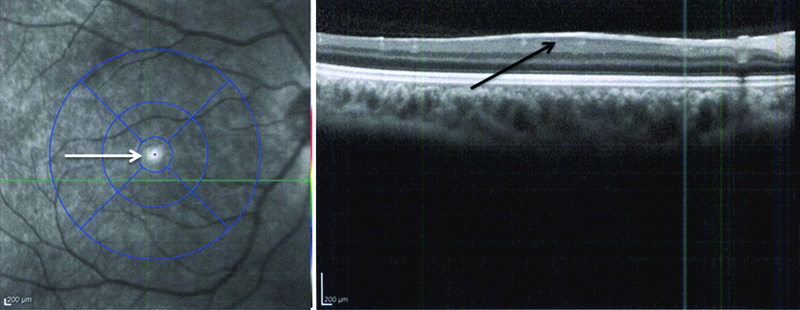
Congenital aniridia manifests as total or partial absence of the iris caused most commonly by mutations in PAX6, FOXC1, PITX2, and CYP1B1. Recently two new genes, FOXD3 and TRIM44, have also been implicated in isolated studies. We discuss the genotype-phenotype correlations for the main implicated genes. Classic aniridia is a panocular condition, which includes aniridia, cataract, corneal pannus, foveal, and optic nerve hypoplasia associated with mutations in the PAX6 gene. Classical aniridia is due to PAX6 mutations, while other genes contribute to aniridia-like phenotypes. We review the challenges involved in the management of aniridia, and discuss various surgical interventions. The clinical importance of defining the genotype in cases of congenital aniridia has become acutely apparent with the advent of possible therapies for classical aniridia, which are discussed.
Keywords: aniridia; aniridia-associated keratopathy; cataract; congenital; goniotomy.
Figures
Similar articles
-
Comprehensive Analysis of Congenital Aniridia and Differential Diagnoses: Genetic Insights and Clinical Manifestations.Ophthalmol Ther. 2025 May;14(5):835-856. doi: 10.1007/s40123-025-01122-1. Epub 2025 Mar 26. Ophthalmol Ther. 2025. PMID: 40138169 Free PMC article. Review.
-
Clinical and molecular aspects of congenital aniridia - A review of current concepts.Indian J Ophthalmol. 2022 Jul;70(7):2280-2292. doi: 10.4103/ijo.IJO_2255_21. Indian J Ophthalmol. 2022. PMID: 35791108 Free PMC article. Review.
-
Mutational analysis and genotype-phenotype correlations in southern Indian patients with sporadic and familial aniridia.Mol Vis. 2015 Jan 27;21:88-97. eCollection 2015. Mol Vis. 2015. PMID: 25678763 Free PMC article.
-
Aniridic Fibrosis Syndrome.2023 Jul 18. In: StatPearls [Internet]. Treasure Island (FL): StatPearls Publishing; 2025 Jan–. 2023 Jul 18. In: StatPearls [Internet]. Treasure Island (FL): StatPearls Publishing; 2025 Jan–. PMID: 32119423 Free Books & Documents.
-
Two Paired Box 6 mutations identified in Chinese patients with classic congenital aniridia and cataract.Mol Med Rep. 2018 Nov;18(5):4439-4445. doi: 10.3892/mmr.2018.9469. Epub 2018 Sep 10. Mol Med Rep. 2018. PMID: 30221735 Free PMC article.
Cited by
-
Systemic Diseases in Patients with Congenital Aniridia: A Report from the Homburg Registry for Congenital Aniridia.Ophthalmol Ther. 2025 Feb;14(2):433-445. doi: 10.1007/s40123-024-01084-w. Epub 2025 Jan 4. Ophthalmol Ther. 2025. PMID: 39755898 Free PMC article.
-
Clinical and Genetic Correlation in Neurocristopathies: Bridging a Precision Medicine Gap.J Clin Med. 2024 Apr 11;13(8):2223. doi: 10.3390/jcm13082223. J Clin Med. 2024. PMID: 38673496 Free PMC article. Review.
-
Comprehensive Analysis of Congenital Aniridia and Differential Diagnoses: Genetic Insights and Clinical Manifestations.Ophthalmol Ther. 2025 May;14(5):835-856. doi: 10.1007/s40123-025-01122-1. Epub 2025 Mar 26. Ophthalmol Ther. 2025. PMID: 40138169 Free PMC article. Review.
-
A rare case of congenital aniridia with an unusual run-on mutation in PAX6 gene.Indian J Ophthalmol. 2022 Jul;70(7):2661-2664. doi: 10.4103/ijo.IJO_433_22. Indian J Ophthalmol. 2022. PMID: 35791194 Free PMC article. No abstract available.
-
Congenital aniridia: clinical profile of children seen at the University College Hospital, Ibadan, South-West Nigeria.Ther Adv Ophthalmol. 2021 May 31;13:25158414211019513. doi: 10.1177/25158414211019513. eCollection 2021 Jan-Dec. Ther Adv Ophthalmol. 2021. PMID: 34104869 Free PMC article.
References
-
- Nelson LB, Spaeth GL, Nowinski TS, Margo CE, and Jackson L (1984). Aniridia. A review. Survey of ophthalmology 28, 621–642. - PubMed
-
- Sharma V, and Mohan M (2010). Traumatic aniridia and self-sealed globe rupture following blunt trauma. Eye 24, 1526. - PubMed
-
-
Grant WM, and Walton DS (1974). Progressive changes in the angle in congenital aniridia, with development of glaucoma. Am J Ophthalmol 78, 842–847.
** First to report the presence of an iris stump through gonioscopy in all cases of congenital aniridia, and extend findings for progressive angle changes in these patients resulting in glaucoma.
-
-
- Shaham O, Menuchin Y, Farhy C, and Ashery-Padan R (2012). Pax6: a multi-level regulator of ocular development. Progress in retinal and eye research 31, 351–376. - PubMed
-
- Walther C, and Gruss P (1991). Pax-6, a murine paired box gene, is expressed in the developing CNS. Development 113, 1435–1449. - PubMed
Grants and funding
LinkOut - more resources
Full Text Sources
Other Literature Sources