. 2018 Aug;9(2):115-134.
doi: 10.3892/mco.2018.1657. Epub 2018 Jun 14.

Cell cycle checkpoint control: The cyclin G1/Mdm2/p53 axis emerges as a strategic target for broad-spectrum cancer gene therapy - A review of molecular mechanisms for oncologists

Affiliations

Cell cycle checkpoint control: The cyclin G1/Mdm2/p53 axis emerges as a strategic target for broad-spectrum cancer gene therapy - A review of molecular mechanisms for oncologists

Erlinda M Gordon et al. Mol Clin Oncol. 2018 Aug.

Abstract

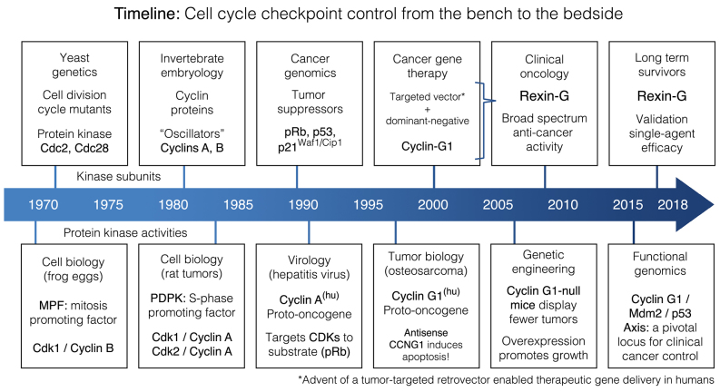
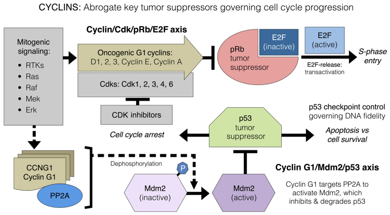
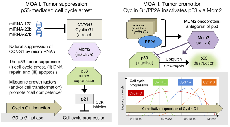
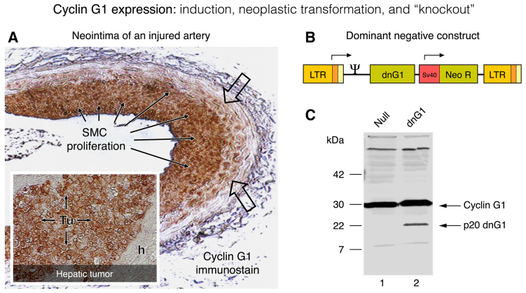
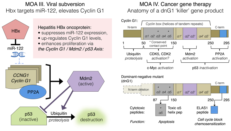
Basic research in genetics, biochemistry and cell biology has identified the executive enzymes and protein kinase activities that regulate the cell division cycle of all eukaryotic organisms, thereby elucidating the importance of site-specific protein phosphorylation events that govern cell cycle progression. Research in cancer genomics and virology has provided meaningful links to mammalian checkpoint control elements with the characterization of growth-promoting proto-oncogenes encoding c-Myc, Mdm2, cyclins A, D1 and G1, and opposing tumor suppressor proteins, such as p53, pRb, p16INK4A and p21WAF1, which are commonly dysregulated in cancer. While progress has been made in identifying numerous enzymes and molecular interactions associated with cell cycle checkpoint control, the marked complexity, particularly the functional redundancy, of these cell cycle control enzymes in mammalian systems, presents a major challenge in discerning an optimal locus for therapeutic intervention in the clinical management of cancer. Recent advances in genetic engineering, functional genomics and clinical oncology converged in identifying cyclin G1 (CCNG1 gene) as a pivotal component of a commanding cyclin G1/Mdm2/p53 axis and a strategic locus for re-establishing cell cycle control by means of therapeutic gene transfer. The purpose of the present study is to provide a focused review of cycle checkpoint control as a practicum for clinical oncologists with an interest in applied molecular medicine. The aim is to present a unifying model that: i) clarifies the function of cyclin G1 in establishing proliferative competence, overriding p53 checkpoints and advancing cell cycle progression; ii) is supported by studies of inhibitory microRNAs linking CCNG1 expression to the mechanisms of carcinogenesis and viral subversion; and iii) provides a mechanistic basis for understanding the broad-spectrum anticancer activity and single-agent efficacy observed with dominant-negative cyclin G1, whose cytocidal mechanism of action triggers programmed cell death. Clinically, the utility of companion diagnostics for cyclin G1 pathways is anticipated in the staging, prognosis and treatment of cancers, including the potential for rational combinatorial therapies.

Keywords: CCNG1; Mdm2; Rexin-G; cancer gene therapy; cyclin-dependent kinase; gene delivery; microRNAs; p53; protein phosphatase 2A; proto-oncogene; tumor suppressor.


Figures

Figure 1.
Figure 1.
Timeline of multidisciplinary milestones in the development of cancer gene therapy.
Figure 2.
Figure 2.
Diagram of oncogenic G1 cyclin functions arrayed in biochemical opposition to the pRb tumor suppressor protein that governs cell cycle progression (cyclin/CDK/Rb/E2F axis); this is distinguishable from the function of cyclin G1, which activates the Mdm2 oncoprotein in its opposition to the p53 tumor suppressor that governs cell fate (cyclin G1/Mdm2/p53 axis). CDK, cyclin-dependent kinase; Rb, retinoblastoma.
Figure 3.
Figure 3.
Highlighting dominant features of the commanding cyclin G1/Mdm2/p53 axis in the mechanisms-of-action (MOA) of tumor suppression (MOA I.; left panel) and tumor promotion (MOA II.; right panel). Note: In contrast to the periodic expression of the classic cyclins (D, E, A and B), cyclin G1 expression is normally induced upon ‘exit’ from quiescence (G0-to-G1 boundary; left panel); however, constitutive expression of cyclin G1 remains constant throughout the cell cycle (bottom right panel).
Figure 4.
Figure 4.
Cyclin G1 expression: Induction, neoplastic transformation and knockout. The characteristic induction of cyclin G1 protein (G0-to-G1 phase) is visualized by immunohistochemistry (A; brown staining) in the activated periphery (open arrows) and the proliferative smooth muscle cells (SMC; solid arrows) in a rat carotid artery model of vascular restenosis following balloon catheter injury. For comparison, the constitutively high levels of cyclin G1 expression seen in a flagrant pancreatic cancer xenograft (Tu), metastatic to the liver (A; insert) is contrasted by the negligible expression of cyclin G1 seen in the adjacent normal (host) nude mouse hepatocytes(h). The design of a dominant-negative (knockout) construct of cyclin G1 (p20 dnG1) is shown in the context of its MoMuLV retroviral expression vector (B); the truncated p20 dnG1 protein (devoid of N-terminal and α1, α2 cyclin box domains) induces apoptosis in the presence of the abundant wild-type cyclin G1, as shown in western blots of cellular proteins (C).
Figure 5.
Figure 5.
Regional delivery of the matrix/lesion targeted vector to the liver via the portal vein. Early metastasis (left plate A-C: A, H&E staining); detection of the β-galactosidase transgene (B and C) in metastatic tumor cells (arrows). Tumor angiogenesis (left plate D-F: D, H&E stain); detection of the β-gal transgene (E and F) in proliferative vascular endothelial cells (arrows). Flagrant tumors (right plate A, C magnified) of untreated control animals is shown in contrast to the dose-dependent Reduction of tumor foci (right plate B, D magnified) achieved by repeated portal vein infusions of the cytocidal Mx-dnG1 vector. Note: The residual immune infiltrates (B arrows, D golden cells magnified) are hemosiderin-laden Kupffer cells engaged in the elimination of residual tumor debris.
Figure 6.
Figure 6.
Left panel: Depicts tumor-targeted vector delivery (A, B magnified vs. C control) and subsequent β-gal transgene expression (D, E magnified vs. F control) following repeated intravenous vector infusions in a subcutaneous xenograft model of metastatic pancreatic cancer. Right panel: Massive apoptosis (arrows: C and D, high mag; E, stroma; F, necrosis) in association with anti-angiogenesis and focal necrosis, compared with control tumors (A, B magnified) where angiogenesis is robust and apoptosis rare.
Figure 7.
Figure 7.
Characteristic changes in tumor histology following intravenous Mx-dnG1 treatment. Untreated tumor xenografts (left panel) exhibit robust tumor (tu) cell proliferation with active zones of neo-angiogenesis (A, open arrows; B, magnified); Masson's trichrome stain (C, D magnified) reveals exposed collagenous proteins (solid arrows, blue stain) associated primarily with angiogenesis and reactive stroma formation (st). By contrast, Mx-dnG1 treatment (right panel) reduces the flagrant population of proliferative tumor cells (tu) significantly (A, boxed; B, magnified), producing large regions of focal necrosis (nec) accompanied by overt anti-angiogenesis (*). Residual tumors are ultimately reduced to besieged clusters of proliferative tumor (tu) cells (C, boxed; D, magnified), as reactive/reparative fibrosis (fib), including nascent (i.e., targetable) ECM deposition (blue stain), dominates the histology of tumor regression observed in Mx-dnG1 treated animals.
Figure 8.
Figure 8.
Histochemical analysis of a tumor biopsy: Pancreatic cancer, metastatic to the liver, following intravenous infusions of tumor-targeted Mx-dnG1 (Rexin-G). Left panel, tumor targeting: Immunohistochemical staining for the retrovector gp70 envelope protein (brown staining) demonstrates widespread vector penetration and accumulation within the tumor (A), with particularly high levels of immunostaining appearing in the cancer cells (A and B, arrows) and associated vasculature (C, arrows). Right panel, mechanisms of action: (A) Clusters of residual (CK17+) pancreatic cancer cells (A, inset of western blot) are distinguished from the reactive fibrosis (fib; A, inset) seen with Masson's trichrome stain, which stains the collagenous extracellular matrix proteins bright blue. Application of the TUNEL method for detecting apoptotic DNA fragmentation reveals massive levels of apoptosis (arrows) in cancer cells (B vs. C, control) as the actual mechanism of dnG1 action.
Figure 9.
Figure 9.
Left panel: Mechanism-of-action (MOA) III. HBx-mediated viral subversion of the hepatocellular division cycle operates by suppression of miR-122, hence de-repression of CCNG1 expression. Right panel: MOA IV. Cancer gene therapy, dng1 structure. The cytocidal dnG1 protein, a dominant-negative mutant construct of cyclin G1, is devoid of the ‘ubiquitinated’ N-terminus (proteolytic processing), as well as the first two helical segments (α1 and α2) of the definitive cyclin box, characteristically arrayed in cyclins as a tandem set of helical segments, including two highly-conserved residues (asterisks) essential for cyclin-dependent kinase (CDK) binding. The cytocidal dnG1 protein, which induces apoptosis in proliferative cells, retains the presumptive (?) CDK contact points (Helix α3*, α5*) and the structural domains attributed to PP2A, β' and Mdm2 binding. Remarkably, new therapeutic peptides (e.g., ELAS1 and α5 Helix peptides) derived from structures or homologous interfaces contained within the dnG1 protein are reported to induce cell cycle blockade and apoptosis, respectively.

References

    1. Goel G, Makkar HP, Francis G, Becker K. Phorbol esters: Structure, biological activity, and toxicity in animals. Int J Toxicol. 2007;26:279–288. doi: 10.1080/10915810701464641. - DOI - PubMed
    1. Weinstein IB. The origins of human cancer: Molecular mechanisms of carcinogenesis and their implications for cancer prevention and treatment-twenty-seventh G.H.A. Clowes memorial award lecture. Cancer Res. 1988;48:4135–4143. - PubMed
    1. Kikkawa U, Takai Y, Tanaka Y, Miyake R, Nishizuka Y. Protein kinase C as a possible receptor protein of tumor-promoting phorbol esters. J Biol Chem. 1983;258:11442–11445. - PubMed
    1. Cooke M, Magimaidas A, Casado-Medrano V, Kazanietz MG. Protein kinase C in cancer: The top five unanswered questions. Mol Carcinog. 2017;56:1531–1542. doi: 10.1002/mc.22617. - DOI - PMC - PubMed
    1. Reid BJ, Culotti JG, Nash RS, Pringle JR. Forty-five years of cell-cycle genetics. Mol Biol Cell. 2015;26:4307–4312. doi: 10.1091/mbc.e14-10-1484. - DOI - PMC - PubMed