

Left lateralized cerebral glucose metabolism declines in amyloid-β positive persons with mild cognitive impairment
- PMID: 30101060
- PMCID: PMC6084012
- DOI: 10.1016/j.nicl.2018.07.016
Left lateralized cerebral glucose metabolism declines in amyloid-β positive persons with mild cognitive impairment
Abstract
Background: Previous publications indicate that Alzheimer's Disease (AD) related cortical atrophy may develop in asymmetric patterns, with accentuation of the left hemisphere. Since fluorodeoxyglucose positron emission tomography (FDG PET) measurements of the regional cerebral metabolic rate of glucose (rCMRgl) provide a sensitive and specific marker of neurodegenerative disease progression, we sought to investigate the longitudinal pattern of rCMRgl in amyloid-positive persons with mild cognitive impairment (MCI) and dementia, hypothesizing asymmetric declines of cerebral glucose metabolism.
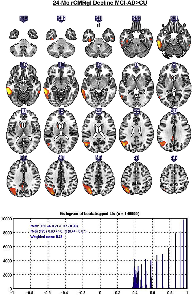
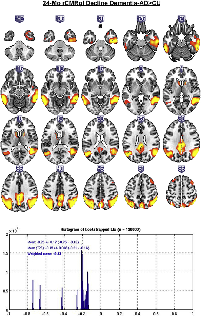
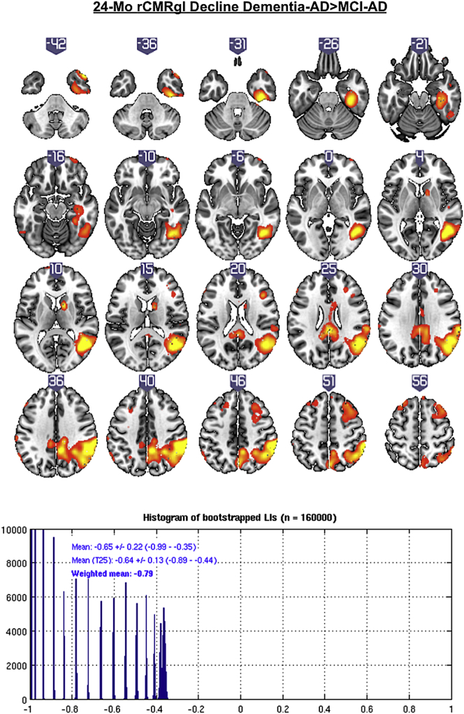
Methods: Using florbetapir PET and cerebrospinal fluid (CSF) measures to define amyloid-β (Aβ) positivity, 40 Aβ negative (Aβ-) cognitively unimpaired controls (CU; 76 ± 5y), 76 Aβ positive (Aβ+) persons with MCI (76 ± 7y) and 51 Aβ + persons with probable AD dementia (75 ± 7y) from the AD Neuroimaging Initiative (ADNI) were included in this study with baseline and 2-year follow-up FDG PET scans. The degree of lateralization of longitudinal rCMRgl declines in subjects with Aβ + MCI and AD in comparison with Aβ- CU were statistically quantified via bootstrapped lateralization indices [(LI); range - 1 (right) to 1 (left)].
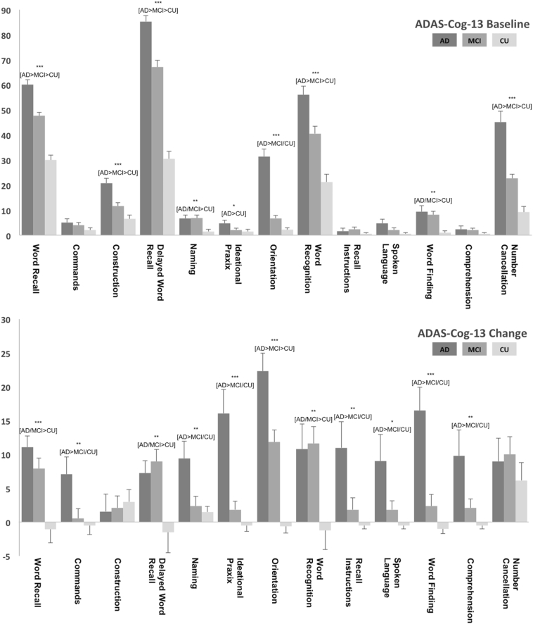
Results: Compared to Aβ- CU, Aβ + MCI patients showed marked left hemispheric lateralization (LI: 0.78). In contrast, modest right hemispheric lateralization (LI: -0.33) of rCMRgl declines was found in Aβ + persons with probable AD dementia. Additional comparisons of Aβ + groups (i.e. MCI and probable AD dementia) consequently indicated right hemispheric lateralization (LI: -0.79) of stronger rCMRgl declines in dementia stages of AD. For all comparisons, voxel-based analyses confirmed significant (pFWE<0.05) declines of rCMRgl within AD-typical brain regions. Analyses of cognitive data yielded predominant decline of memory functions in both MCI and dementia stages of AD.
Conclusions: These data indicate that in early stages, AD may be characterized by a more lateralized pattern of left hemispheric rCMRgl declines. However, metabolic differences between hemispheres appear to diminish with further progression of the disease.
Keywords: Alzheimer's disease; FDG PET; Lateralization; MCI.
Figures
References
-
- Buckner R.L., Snyder A.Z., Shannon B.J., Larossa G., Sachs R., Fotenos A.F., Sheline Y.I., Klunk W.E., Mathis C.A., Morris J.C., Mintun M.A. Molecular, structural, and functional characterization of Alzheimer's disease: evidence for a relationship between default activity, amyloid, and memory. J. Neurosci. 2005;25:7709–7717. - PMC - PubMed
-
- De Winter F.-L., Zhu Q., Van den Stock J., Nelissen K., Peeters R., de Gelder B., Vanduffel W., Vandenbulcke M. Lateralization for dynamic facial expressions in human superior temporal sulcus. NeuroImage. 2015;106:340–352. - PubMed
Publication types
MeSH terms
Substances
Grants and funding
LinkOut - more resources
Full Text Sources
Other Literature Sources
Medical
