

Sex-specific hippocampus volume changes in obstructive sleep apnea
- PMID: 30101062
- PMCID: PMC6083433
- DOI: 10.1016/j.nicl.2018.07.027
Sex-specific hippocampus volume changes in obstructive sleep apnea
Abstract
Introduction: Obstructive sleep apnea (OSA) patients show hippocampal-related autonomic and neurological symptoms, including impaired memory and depression, which differ by sex, and are mediated in distinct hippocampal subfields. Determining sites and extent of hippocampal sub-regional injury in OSA could reveal localized structural damage linked with OSA symptoms.
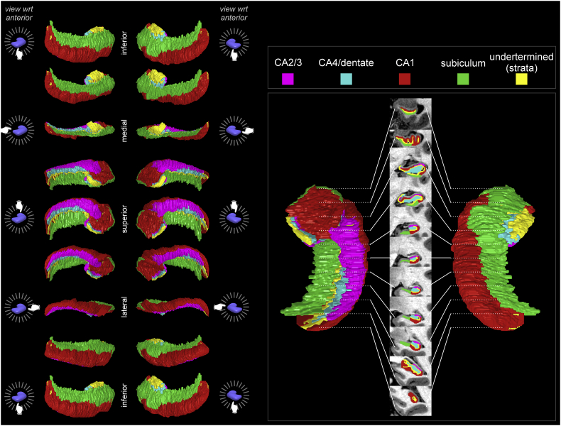
Methods: High-resolution T1-weighted images were collected from 66 newly-diagnosed, untreated OSA (mean age ± SD: 46.3 ± 8.8 years; mean AHI ± SD: 34.1 ± 21.5 events/h;50 male) and 59 healthy age-matched control (46.8 ± 9.0 years;38 male) participants. We added age-matched controls with T1-weighted scans from two datasets (IXI, OASIS-MRI), for 979 controls total (426 male/46.5 ± 9.9 years). We segmented the hippocampus and analyzed surface structure with "FSL FIRST" software, scaling volumes for brain size, and evaluated group differences with ANCOVA (covariates: total-intracranial-volume, sex; P < .05, corrected).
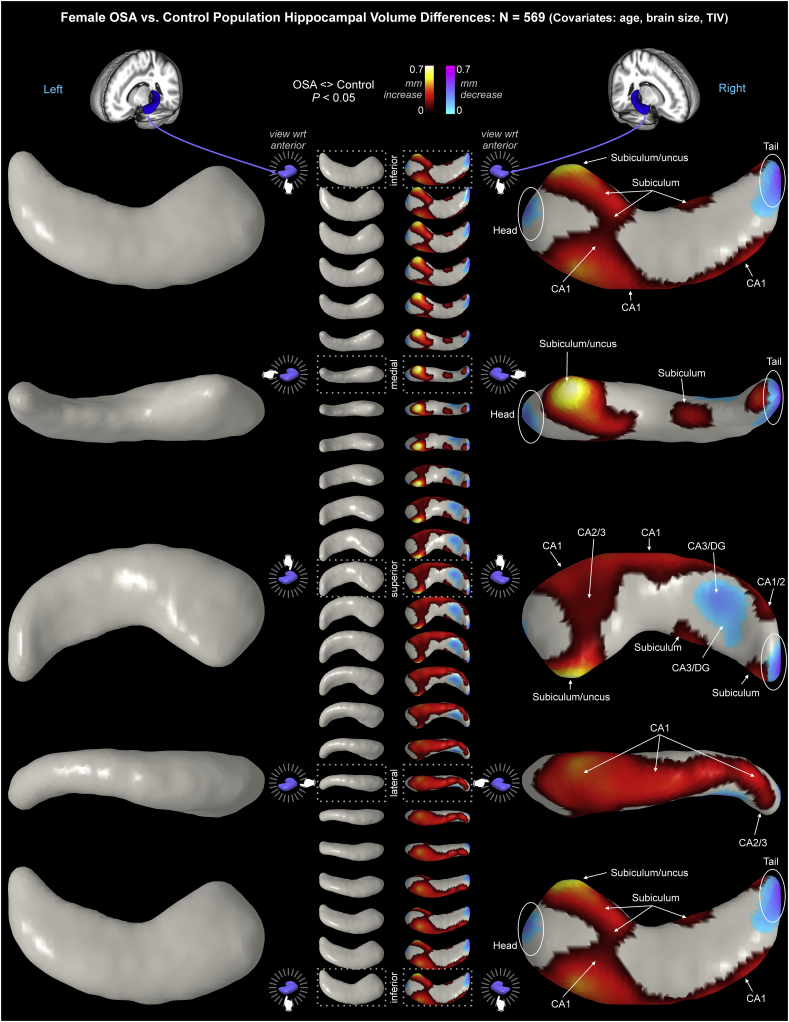
Results: In OSA relative to controls, the hippocampus showed small areas larger volume bilaterally in CA1 (surface displacement ≤0.56 mm), subiculum, and uncus, and smaller volume in right posterior CA3/dentate (≥ - 0.23 mm). OSA vs. control males showed higher bilateral volume (≤0.61 mm) throughout CA1 and subiculum, extending to head and tail, with greater right-sided increases; lower bilateral volumes (≥ - 0.45 mm) appeared in mid- and posterior-CA3/dentate. OSA vs control females showed only right-sided effects, with increased CA1 and subiculum/uncus volumes (≤0.67 mm), and decreased posterior CA3/dentate volumes (≥ - 0.52 mm). Unlike males, OSA females showed volume decreases in the right hippocampus head and tail.
Conclusions: The hippocampus shows lateralized and sex-specific, OSA-related regional volume differences, which may contribute to sex-related expression of symptoms in the sleep disorder. Volume increases suggest inflammation and glial activation, whereas volume decreases suggest long-lasting neuronal injury; both processes may contribute to dysfunction in OSA.
Keywords: AHI, apnea-hypopnea index; Autonomic; CA, cornu ammonis; Inflammation; Intermittent hypoxia; Neuroimaging; OSA, obstructive sleep apnea; Oxidative stress.
Figures


References
-
- Alchanatis M., Deligiorgis N., Zias N., Amfilochiou A., Gotsis E., Karakatsani A. Frontal brain lobe impairment in obstructive sleep apnoea: a proton MR spectroscopy study. Eur. Respir. J. 2004;24:980–986. - PubMed
-
- Algin O., Gokalp G., Ocakoglu G., Ursavas A., Taskapilioglu O., Hakyemez B. Neurochemical-structural changes evaluation of brain in patients with obstructive sleep apnea syndrome. Eur. J. Radiol. 2012;81:491–495. - PubMed
-
- Alkan A., Sharifov R., Akkoyunlu M.E., Kilicarslan R., Toprak H., Aralasmak A. MR spectroscopy features of brain in patients with mild and severe obstructive sleep apnea syndrome. Clin. Imaging. 2013;37:989–992. - PubMed
-
- Author Sleep-related breathing disorders in adults: recommendations for syndrome definition and measurement techniques in clinical research. The Report of an American Academy of Sleep Medicine Task Force. Sleep. 1999;22:667–689. - PubMed
Publication types
MeSH terms
Grants and funding
LinkOut - more resources
Full Text Sources
Other Literature Sources
Medical
Miscellaneous
