

Surgical treatment of an umbilical hernia in a free-ranging sub-adult African elephant in Samburu National Reserve, Kenya
- PMID: 30101103
- PMCID: PMC6070018
- DOI: 10.2147/VMRR.S74756
Surgical treatment of an umbilical hernia in a free-ranging sub-adult African elephant in Samburu National Reserve, Kenya
Abstract
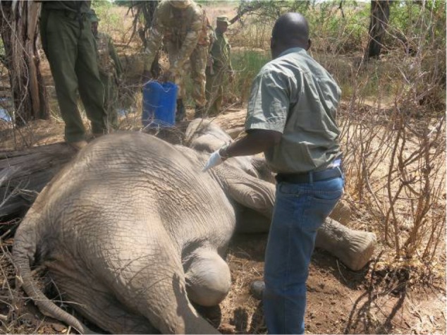
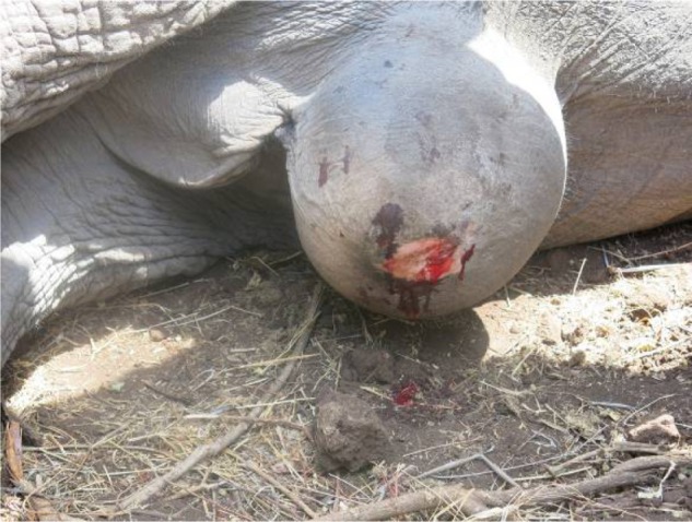
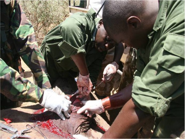
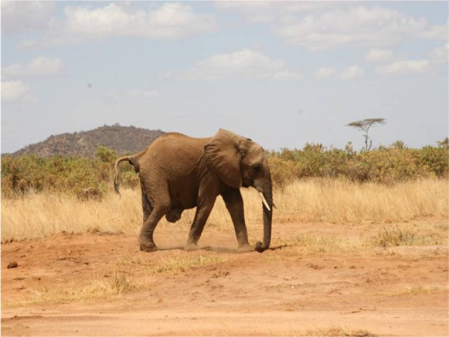
A 10-year-old male African elephant (Loxodonta africana) at Samburu National Reserve in Northern Kenya, weighing approximately 1,600 kg, presented with an umbilical hernia in October 2013. Umbilical herniorrhaphy was carried out under field conditions. Anesthesia was induced and maintained using etorphine hydrochloride for 3 hours during the surgery. This case report details both the surgical and anesthetic procedure carried out to correct the hernia, and the eventual successful recovery of the elephant from anesthesia. However, the elephant died weeks after the surgery and a postmortem could not reveal the cause of death because predators had scavenged the carcass. The challenges of the surgical procedure and outcome including possible causes of death are highlighted in this report.
Keywords: African elephant; etorphine hydrochloride; general anesthesia; local anesthesia Lignocaine + adrenaline; umbilical herniorrhaphy.
Conflict of interest statement
Disclosure The authors report no conflicts of interest in this work.
Figures
References
-
- Loxodonta africana. Blanc J; 2008. [Accessed September 10, 2014]. International Union for Conservation of Nature [homepage on the Internet] (The IUCN Red List of Threatened Species). Version 2014.2. Available from: http://www.iucnredlist.org.
-
- Singh BS. Umbilical hernia in an elephant calf. Indian Vet J. 1971;48(5):533–536. - PubMed
-
- Fowler ME. Problems with immobilizing and anesthetizing elephants. Proc Am Ass Zoo Vet. 1981:87–91.
-
- Oosterhuis JE. The performance of caesarian section on an Asian elephant (Elephas maximus indicus) Proc Am Zoo Vet. 1990:157–158.
-
- Pathak S, Saikia CJ, Lahon DK, et al. Attempted ventral herniorrhaphy in an Asian elephant (Elephas maximus) using xylaxine sedation. J Zoo Wildl Med. 1990;21:234–235.
Publication types
LinkOut - more resources
Full Text Sources
