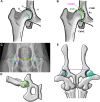
Diagnosis, prevention, and management of canine hip dysplasia: a review
- PMID: 30101105
- PMCID: PMC6070021
- DOI: 10.2147/VMRR.S53266
Diagnosis, prevention, and management of canine hip dysplasia: a review
Abstract
Canine hip dysplasia (CHD) is a polygenic and multifactorial developmental disorder characterized by coxofemoral (hip) joint laxity, degeneration, and osteoarthritis (OA). Current diagnostic techniques are largely subjective measures of joint conformation performed at different stages of development. Recently, measures on three-dimensional images generated from computed tomography scans predicted the development of OA associated with CHD. Continued refinement of similar imaging methods may improve diagnostic imaging techniques to identify dogs predisposed to degenerative hip joint changes. By current consensus, joint changes consistent with CHD are influenced by genetic predisposition as well as environmental and biomechanical factors; however, despite decades of work, the relative contributions of each to the development and extent of CHD signs remain elusive. Similarly, despite considerable effort to decipher the genetic underpinnings of CHD for selective breeding programs, relevant genetic loci remain equivocal. As such, prevention of CHD within domestic canine populations is marginally successful. Conservative management is often employed to manage signs of CHD, with lifelong maintenance of body mass as one of the most promising methods. Surgical intervention is often employed to prevent joint changes or restore joint function, but there are no gold standards for either goal. To date, all CHD phenotypes are considered as a single entity in spite of recognized differences in expression and response to environmental conditions and treatment. Identification of distinct CHD phenotypes and targeting evidence-based conservative and invasive treatments for each may significantly advance prevention and management of a prevalent, debilitating condition in canine companions.
Keywords: canine hip dysplasia; joint; orthopedics; osteoarthritis.
Conflict of interest statement
Disclosure The authors report no conflicts of interest in this work.
Figures


Similar articles
-
Emerging insights into the genetic basis of canine hip dysplasia.Vet Med (Auckl). 2015 May 20;6:193-202. doi: 10.2147/VMRR.S63536. eCollection 2015. Vet Med (Auckl). 2015. PMID: 30101106 Free PMC article. Review.
-
Canine hip dysplasia: phenotypic scoring and the role of estimated breeding value analysis.N Z Vet J. 2015 Mar;63(2):69-78. doi: 10.1080/00480169.2014.949893. Epub 2014 Dec 1. N Z Vet J. 2015. PMID: 25072401
-
Relationships of hip joint volume ratios with degrees of joint laxity and degenerative disease from youth to maturity in a canine population predisposed to hip joint osteoarthritis.Am J Vet Res. 2011 Mar;72(3):376-83. doi: 10.2460/ajvr.72.3.376. Am J Vet Res. 2011. PMID: 21355741 Free PMC article.
-
Chronology of hip dysplasia development in a cohort of 48 Labrador retrievers followed for life.Vet Surg. 2012 Jan;41(1):20-33. doi: 10.1111/j.1532-950X.2011.00935.x. Vet Surg. 2012. PMID: 23253036
-
Diagnosis, genetic control and preventive management of canine hip dysplasia: a review.Vet J. 2010 Jun;184(3):269-76. doi: 10.1016/j.tvjl.2009.04.009. Epub 2009 May 9. Vet J. 2010. PMID: 19428274 Review.
Cited by
-
B-mode ultrasonography and ARFI elastography of articular and peri-articular structures of the hip joint in non-dysplastic and dysplastic dogs as confirmed by radiographic examination.BMC Vet Res. 2023 Oct 2;19(1):181. doi: 10.1186/s12917-023-03753-7. BMC Vet Res. 2023. PMID: 37784120 Free PMC article.
-
Long-Term Effects of Whole-Body Vibration on Hind Limb Muscles, Gait and Pain in Lame Dogs with Borderline-to-Severe Hip Dysplasia-A Pilot Study.Animals (Basel). 2023 Nov 9;13(22):3456. doi: 10.3390/ani13223456. Animals (Basel). 2023. PMID: 38003074 Free PMC article.
-
GenPup-M: A novel validated owner-reported clinical metrology instrument for detecting early mobility changes in dogs.PLoS One. 2023 Dec 27;18(12):e0291035. doi: 10.1371/journal.pone.0291035. eCollection 2023. PLoS One. 2023. PMID: 38150469 Free PMC article.
-
Stratification of Companion Animal Life Stages from Electronic Medical Record Diagnosis Data.J Gerontol A Biol Sci Med Sci. 2023 Mar 30;78(4):579-586. doi: 10.1093/gerona/glac220. J Gerontol A Biol Sci Med Sci. 2023. PMID: 36330848 Free PMC article.
-
Ultrasonographic Ventral Hip Joint Approach and Relationship with Joint Laxity in Estrela Mountain Dogs.Animals (Basel). 2025 Feb 13;15(4):547. doi: 10.3390/ani15040547. Animals (Basel). 2025. PMID: 40003029 Free PMC article.
References
-
- Lust G. An overview of the pathogenesis of canine hip dysplasia. J Am Vet Med Assoc. 1997;210:1443–1445. - PubMed
-
- Zhang ZW, Zhu L, Sandler J, et al. Estimation of heritabilities, genetic correlations, and breeding values of four traits that collectively define hip dysplasia in dogs. Am J Vet Res. 2009;70:483–492. - PubMed
-
- Schnelle GB. Bilateral congenital subluxation of the coxo-femoral joints in a dog. University of Pennsylvania Bulletin School of Veterinary Medicine Veterinary Extension Quarterly. 1937;37:15–16.
-
- Impellizeri JA, Tetrick MA, Muir P. Effect of weight reduction on clinical signs of lameness in dogs with hip osteoarthritis. J Am Vet Med Assoc. 2001;216:1089–1091. - PubMed
-
- Kealy RD, Lawler DF, Ballam JM, et al. Evaluation of the effect of limited food consumption on radiographic evidence of osteoarthritis in dogs. J Am Vet Med Assoc. 2000;217:1678–1680. - PubMed
Publication types
LinkOut - more resources
Full Text Sources
Research Materials

