Case Reports
. 2018 Dec 15;57(24):3529-3535.
doi: 10.2169/internalmedicine.0294-17. Epub 2018 Aug 10.

Pancreatic Acinar Cell Carcinoma with Multiple Liver Metastases Effectively Treated by S-1 Chemotherapy

Affiliations
Case Reports

Pancreatic Acinar Cell Carcinoma with Multiple Liver Metastases Effectively Treated by S-1 Chemotherapy

Naoki Yoshida et al. Intern Med. .

Abstract

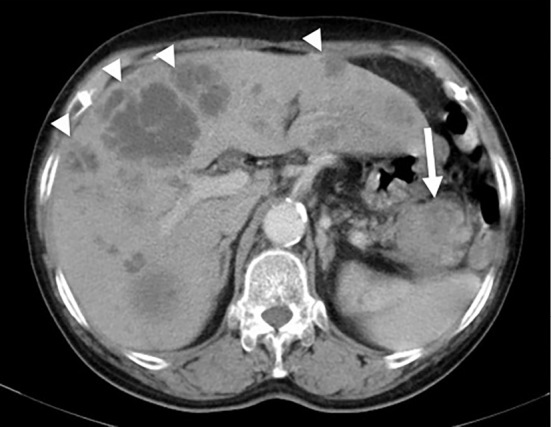
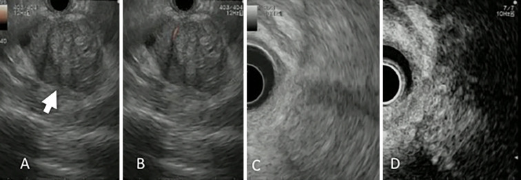
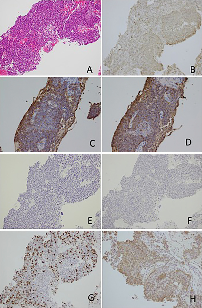
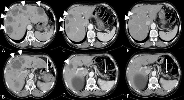
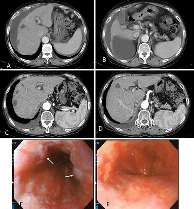
A 79-year-old woman was referred for pancreatic tail cancer with multiple liver metastases. The pancreatic tail tumor was diagnosed as acinar cell carcinoma (ACC) histologically by endoscopic ultrasound-guided fine-needle aspiration. Because of multiple liver metastases, S-1 chemotherapy was administered, resulting in a partial response to chemotherapy one year later. After approximately three years, liver atrophy and esophageal varices developed. We suspected S-1 as the cause of the liver cirrhosis. S-1 cessation minimized ascites and improved the esophageal varices. Although S-1 can potentially treat ACC, we should be watchful for liver cirrhosis caused by its long-term administration.

Keywords: acinar; chemotherapy; liver cirrhosis.


Figures

Figure 1.
Figure 1.
Contrast-enhanced abdominal CT performed at the initial examination. Multiple masses with poor contrast enhancement were observed in the liver (arrowhead), and a mass with poor contrast enhancement and an indistinct border was observed in the pancreatic tail (arrow).
Figure 2.
Figure 2.
Endoscopic ultrasound findings. A tumor (arrow) with a relatively distinct border and heterogeneous echo pattern was observed using endoscopic ultrasound (A). Using power Doppler (B) and ultrasound contrast medium (D), we confirmed blood flow within the tumor, indicating a viable tumor. No clear cystic degeneration was observed. C is a referential image of contrast-enhanced image.
Figure 3.
Figure 3.
Histopathological findings of the endoscopic ultrasound fine-needle aspiration sample. Tumor cells with relatively small oval nuclei, eosinophilic cytoplasm, and a solid growth pattern were detected using Hematoxylin and Eosin staining. (A) Positive staining for α1-antichimotrypsin (B), trypsin (C), and Bcl 10 (D). Negative staining for chromogranin A and synaptophysin. (E, F) Approximately 30-40% of cells were positive for Ki67. (G) ×200 magnification. Positive staining for thymidylate synthase (H).
Figure 4.
Figure 4.
S-1 chemotherapy induced changes in the tumor. Prior to starting treatment (A, B), pancreatic tail tumor (arrow) and multiple liver metastases (arrowhead) were seen. Six months after treatment (C, D), marked reductions in both the pancreatic tail tumor and liver metastases were seen. One year after treatment (E, F), the primary lesion had completely disappeared.
Figure 5.
Figure 5.
After three years of treatment, CT revealed increased ascites and liver atrophy (A, B). After four years of treatment, cessation of S-1 minimized the amount of ascites (C, D). After three years of treatment, upper gastrointestinal endoscopy showed varices with red color sign (arrow) in the middle and lower esophagus (E). Cessation of S-1 improved the varices after another year (F).
Figure 6.
Figure 6.
The main metabolic pathway and mechanistic overview of S-1/5-FU. Tegafur is a precursor to 5-FU, gimeracil inhibits the 5-FU catabolic enzyme dihydropyrimidine dehydrogenase (DPD), and oteracil potassium acts in the gastrointestinal tract primarily blocking orotate phosphoribosyltransferase (OPRT) and reducing gastrointestinal mucous membrane disorder. Thymidine phosphorylase, DPD, thymidylate synthase (TS), and OPRT are enzymes required for metabolizing and activating 5-FU. In particular, TS, a rate-limiting enzyme in the DNA synthesis pathway, is important for determining resistance to fluoropyrimidine. Fluorodeoxyuridylate, the 5-FU active metabolite, binds to TS, which inhibits new DNA synthesis and thereby exerts antitumor effects.

Similar articles

Cited by

References

    1. Morohoshi T, Held G, Klöppel G. Exocrine pancreatic tumours and their histological classification. A study based on 167 autopsy and 97 surgical cases. Histopathology 7: 645-661, 1983. - PubMed
    1. La Rosa S, Adsay V, Albarello L, et al. . Clinicopathologic study of 62 acinar cell carcinomas of the pancreas: insights into the morphology and immunophenotype and search for prognostic markers. Am J Surg Pathol 36: 1782-1795, 2012. - PubMed
    1. Kitagami H, Kondo S, Hirano S, Kawakami H, Egawa S, Tanaka M. Acinar cell carcinoma of the pancreas: clinical analysis of 115 patients from Pancreatic Cancer Registry of Japan Pancreas Society. Pancreas 35: 42-46, 2007. - PubMed
    1. Butturini G, Pisano M, Scarpa A, D’Onofrio M, Auriemma A, Bassi C. Aggressive approach to acinar cell carcinoma of the pancreas: a single-institution experience and a literature review. Langenbecks Arch Surg 396: 363-369, 2011. - PubMed
    1. Fitzgerald PJ, Fortner JG, Watson RC, et al. . The value of diagnostic aids in detecting pancreas cancer. Cancer 41: 868-879, 1978. - PubMed

Publication types

MeSH terms