

A Mass Filling the Right Atrium: Primary Cardiac Rhabdomyosarcoma
- PMID: 30101906
- PMCID: PMC6355423
- DOI: 10.2169/internalmedicine.0657-17
A Mass Filling the Right Atrium: Primary Cardiac Rhabdomyosarcoma
Abstract
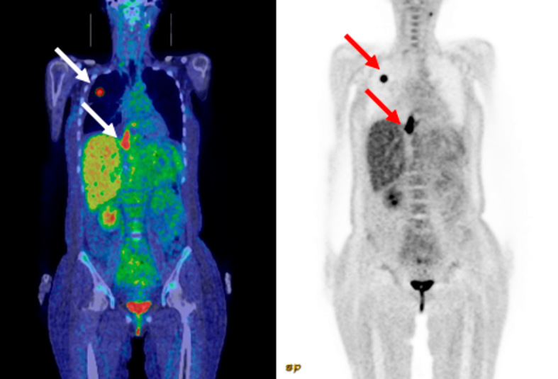
A 43-year-old woman presented with worsening shortness of breath and lower-extremity edema. Echocardiography and computed tomography showed obstruction of blood flow due to a mass filling the right atrium. Emergency surgery was performed for circulatory failure. Primary cardiac rhabdomyosarcoma was diagnosed based on a histological examination. The patient died about two months after the diagnosis despite surgical excision and radiation therapy. The poor prognosis may have resulted from the grossly incomplete removal of the tumor and chemotherapy intolerance. We herein report a case of primary cardiac rhabdomyosarcoma filling the right atrium and offer possible reasons for the poor prognosis.
Keywords: combined modality approach; primary cardiac tumor; prognosis; rhabdomyosarcoma.
Figures


References
-
- McAllister HA, Hall RJ, Cooley DA. Tumors of the heart and pericardium. Curr Probl Cardiol 24: 59-116, 1999. - PubMed
-
- Bouzas-Mosquera A, Flores-Rios X, Aldama G. Primary cardiac rhabdomyosarcoma causing obstruction to the right ventricular outflow. Eur J Echocardiogr 8: 406-407, 2007. - PubMed
-
- Orlandi A, Ferlosio A, Roselli M, Chiariello L, Spagnoli LG. Cardiac sarcomas: an update. J Thorac Oncol 5: 1483-1489, 2010. - PubMed
-
- Simsek H, Sahin M, Gumrukcuoglu HA, Tuncer M, Gunes Y. Recurrence of primary cardiac rhabdomyosarcoma without methastasis two years after surgery. Eur J Gen Med 9: 146-148, 2012.
Publication types
MeSH terms
LinkOut - more resources
Full Text Sources
Other Literature Sources