

CT-guided Biopsy for the Diagnosis of Pulmonary Epithelioid Hemangioendothelioma Mimicking Metastatic Lung Cancer
- PMID: 30101918
- PMCID: PMC6355408
- DOI: 10.2169/internalmedicine.1063-18
CT-guided Biopsy for the Diagnosis of Pulmonary Epithelioid Hemangioendothelioma Mimicking Metastatic Lung Cancer
Abstract
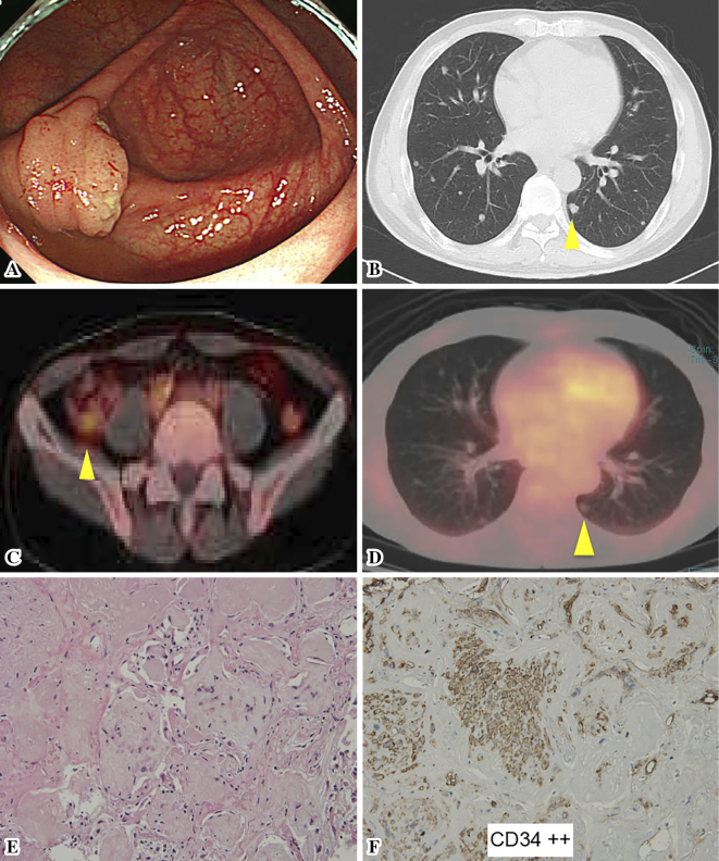
A 69-year-old male patient presented with multiple lung nodules revealed by chest-computed tomography (CT) during a preoperative examination for an appendiceal tumor. The nodule diameters ranged from 2-10 mm without either pleural thickening or effusions. A fluorine-18-labeled fluorodeoxyglucose (18F-FDG)-positron emission tomography (PET)/CT scan showed a high FDG uptake in the appendiceal tumor, but almost normal standardized uptake values in the bilateral lung nodules. A CT-guided biopsy led to a diagnosis of pulmonary epithelioid hemangioendothelioma, a rare vascular tumor with a radiological presentation similar to that of a metastatic lung tumor. The present case is the first to describe successful treatment using a CT-guided biopsy instead of more conventional methods.
Keywords: minimally invasive procedure; multiple lung nodules; thoracoscopic biopsy; vascular tumor.
Figures
Similar articles
-
18F-FDG-PET/CT as an indicator for resection of pulmonary epithelioid hemangioendothelioma.Ann Nucl Med. 2008 Jul;22(6):521-4. doi: 10.1007/s12149-007-0159-z. Epub 2008 Aug 1. Ann Nucl Med. 2008. PMID: 18670859
-
Pulmonary epithelioid hemangioendothelioma.Gen Thorac Cardiovasc Surg. 2011 Apr;59(4):297-300. doi: 10.1007/s11748-010-0651-6. Epub 2011 Apr 12. Gen Thorac Cardiovasc Surg. 2011. PMID: 21484560 Review.
-
Pulmonary epithelioid hemangioendothelioma misdiagnosed as a benign nodule.World J Surg Oncol. 2015 Mar 14;13:107. doi: 10.1186/s12957-015-0518-5. World J Surg Oncol. 2015. PMID: 25889253 Free PMC article.
-
Assessment of indeterminate pulmonary nodules detected in lung cancer screening: Diagnostic accuracy of FDG PET/CT.Lung Cancer. 2016 Jul;97:81-6. doi: 10.1016/j.lungcan.2016.04.025. Epub 2016 May 2. Lung Cancer. 2016. PMID: 27237032
-
Right atrial epithelioid angiosarcoma with multiple pulmonary metastasis confirmed by multimodality imaging-guided pulmonary biopsy: A case report and literature review.Medicine (Baltimore). 2018 Jul;97(30):e11588. doi: 10.1097/MD.0000000000011588. Medicine (Baltimore). 2018. PMID: 30045289 Free PMC article. Review.
Cited by
-
Pleural Epithelioid Hemangioendothelioma (EHE): A Case Report.Cureus. 2023 Jul 3;15(7):e41308. doi: 10.7759/cureus.41308. eCollection 2023 Jul. Cureus. 2023. PMID: 37539424 Free PMC article.
-
Imaging features and deep learning for prediction of pulmonary epithelioid hemangioendothelioma in CT images.J Thorac Dis. 2024 Feb 29;16(2):935-947. doi: 10.21037/jtd-23-455. Epub 2024 Feb 23. J Thorac Dis. 2024. PMID: 38505025 Free PMC article.
-
Primary pulmonary epithelioid hemangioendothelioma.BMJ Case Rep. 2023 Sep 14;16(9):e254915. doi: 10.1136/bcr-2023-254915. BMJ Case Rep. 2023. PMID: 37709495
-
An extremely rare case of pulmonary epithelioid hemangioendothelioma.Thorac Cancer. 2023 Aug;14(24):2519-2522. doi: 10.1111/1759-7714.15051. Epub 2023 Jul 24. Thorac Cancer. 2023. PMID: 37488675 Free PMC article. Review.
References
-
- Watanabe S, Yano F, Kita T, et al. . 18F-FDG-PET/CT as an indicator for resection of pulmonary epithelioid hemangioendothelioma. Ann Nucl Med 22: 521-524, 2008. - PubMed
-
- Weiss SW, Enzinger FM. Epithelioid hemangioendothelioma: a vascular tumor often mistaken for a carcinoma. Cancer 50: 970-981, 1982. - PubMed
-
- Dail DH, Liebow AA. Intravascular bronchioloalveolar tumor. Am J Pathol 78: 6a-7a, 1975.
Publication types
MeSH terms
Substances
LinkOut - more resources
Full Text Sources
Other Literature Sources
Medical
Molecular Biology Databases