

Fibrolamellar Hepatocellular Carcinoma with Multiple Lung Metastases Treated with Multidisciplinary Therapy
- PMID: 30101933
- PMCID: PMC6355421
- DOI: 10.2169/internalmedicine.1243-18
Fibrolamellar Hepatocellular Carcinoma with Multiple Lung Metastases Treated with Multidisciplinary Therapy
Abstract
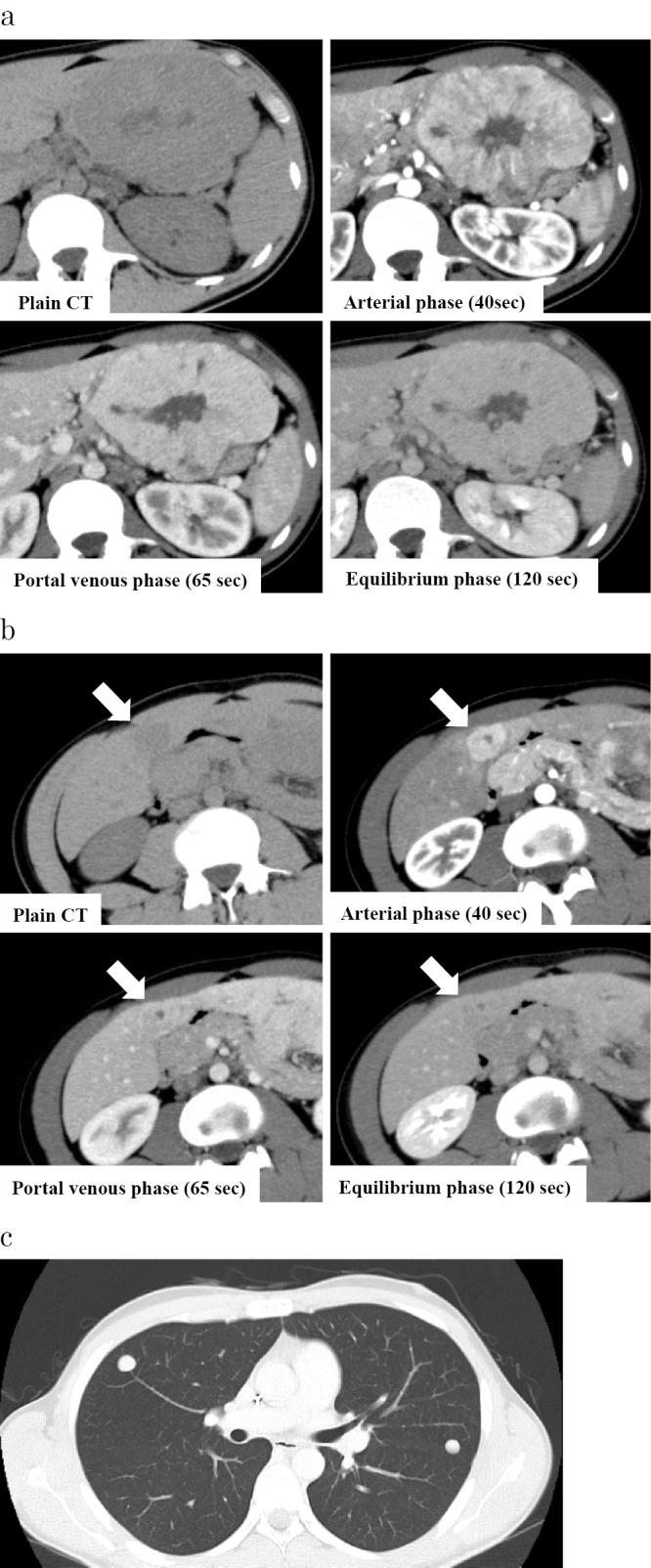
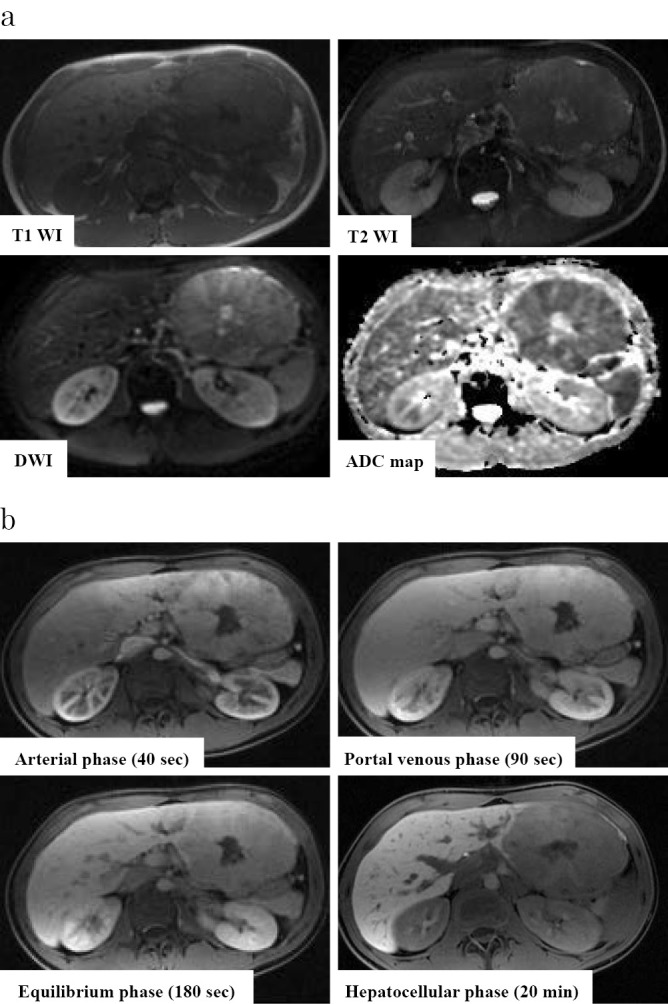
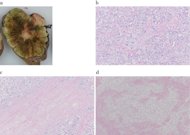
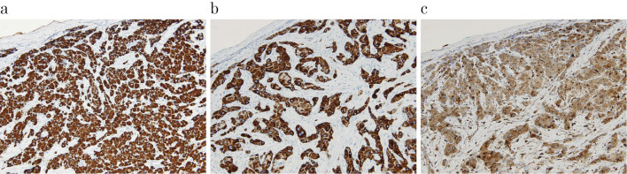
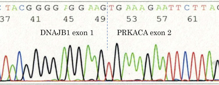
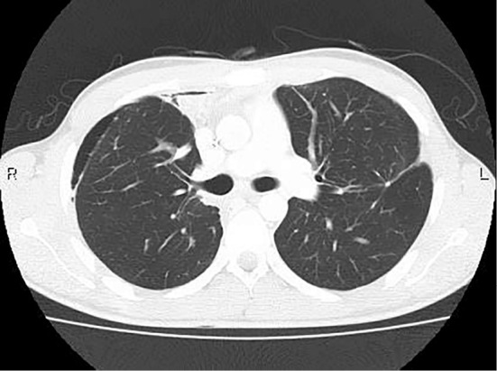
A 20-year old man was diagnosed with fibrolamellar hepatocellular carcinoma (FLHCC) with multiple lung metastases, and chemotherapy with FOLFOX was administered. Contrast enhanced CT after 3 cycles of FOLFOX showed no disease progression. We therefore performed surgical resection and radiofrequency ablation of the liver lesions and lung metastases, after obtaining the patient's informed consent. The liver lesions and lung metastases tested positive for DNAJB1-PRKACA. The treatment for FLHCC with extrahepatic metastasis has not been established; however, in a few cases, good long-term prognoses were obtained with multidisciplinary therapy. We herein report a case of FLHCC with multiple lung metastases that was treated with multidisciplinary therapies.
Keywords: DNAJB1-PRKACA; fibrolamellar hepatocellular carcinoma; multidisciplinary therapy.
Figures
Similar articles
-
[Relapse-Free Survival Following Multidisciplinary Therapy for Hepatocellular Carcinoma with Multiple Lung Metastases--A Case Report].Gan To Kagaku Ryoho. 2015 Nov;42(12):1881-3. Gan To Kagaku Ryoho. 2015. PMID: 26805204 Japanese.
-
Successful multimodal treatment for aggressive metastatic and recurrent fibrolamellar hepatocellular carcinoma in a child.J Pediatr Hematol Oncol. 2014 Jul;36(5):e328-32. doi: 10.1097/MPH.0000000000000137. J Pediatr Hematol Oncol. 2014. PMID: 24608073
-
Percutaneous radiofrequency ablation combined with previous bronchial arterial chemoembolization and followed by radiation therapy for pulmonary metastasis from hepatocellular carcinoma.J Vasc Interv Radiol. 2006 Jul;17(7):1189-93. doi: 10.1097/01.RVI.0000228370.09886.66. J Vasc Interv Radiol. 2006. PMID: 16868173
-
Long-term complete response of advanced hepatocellular carcinoma treated with multidisciplinary therapy including reduced dose of sorafenib: case report and review of the literature.World J Surg Oncol. 2015 Apr 9;13:144. doi: 10.1186/s12957-015-0559-9. World J Surg Oncol. 2015. PMID: 25889667 Free PMC article. Review.
-
Case report: fibrolamellar hepatocellular carcinoma in a Japanese woman: a case report and review of Japanese cases.J Gastroenterol Hepatol. 1996 Jun;11(6):551-5. doi: 10.1111/j.1440-1746.1996.tb01701.x. J Gastroenterol Hepatol. 1996. PMID: 8792309 Review.
Cited by
-
Knockdown of interferon-stimulated gene 15 affects the sensitivity of hepatocellular carcinoma cells to norcantharidin.Exp Ther Med. 2019 Nov;18(5):3751-3758. doi: 10.3892/etm.2019.8028. Epub 2019 Sep 19. Exp Ther Med. 2019. PMID: 31611931 Free PMC article.
References
-
- El-Serag HB, Davila JA. Is fibrolamellar carcinoma different from hepatocellular carcinoma? A US population-based study. Hepatology 39: 798-803, 2004. - PubMed
Publication types
MeSH terms
Supplementary concepts
LinkOut - more resources
Full Text Sources
Other Literature Sources
Medical
Miscellaneous