. 2018 Oct;36(9):847-856.
doi: 10.1038/nbt.4195. Epub 2018 Aug 13.

Targeted delivery of a PD-1-blocking scFv by CAR-T cells enhances anti-tumor efficacy in vivo

Affiliations

Targeted delivery of a PD-1-blocking scFv by CAR-T cells enhances anti-tumor efficacy in vivo

Sarwish Rafiq et al. Nat Biotechnol. 2018 Oct.

Abstract

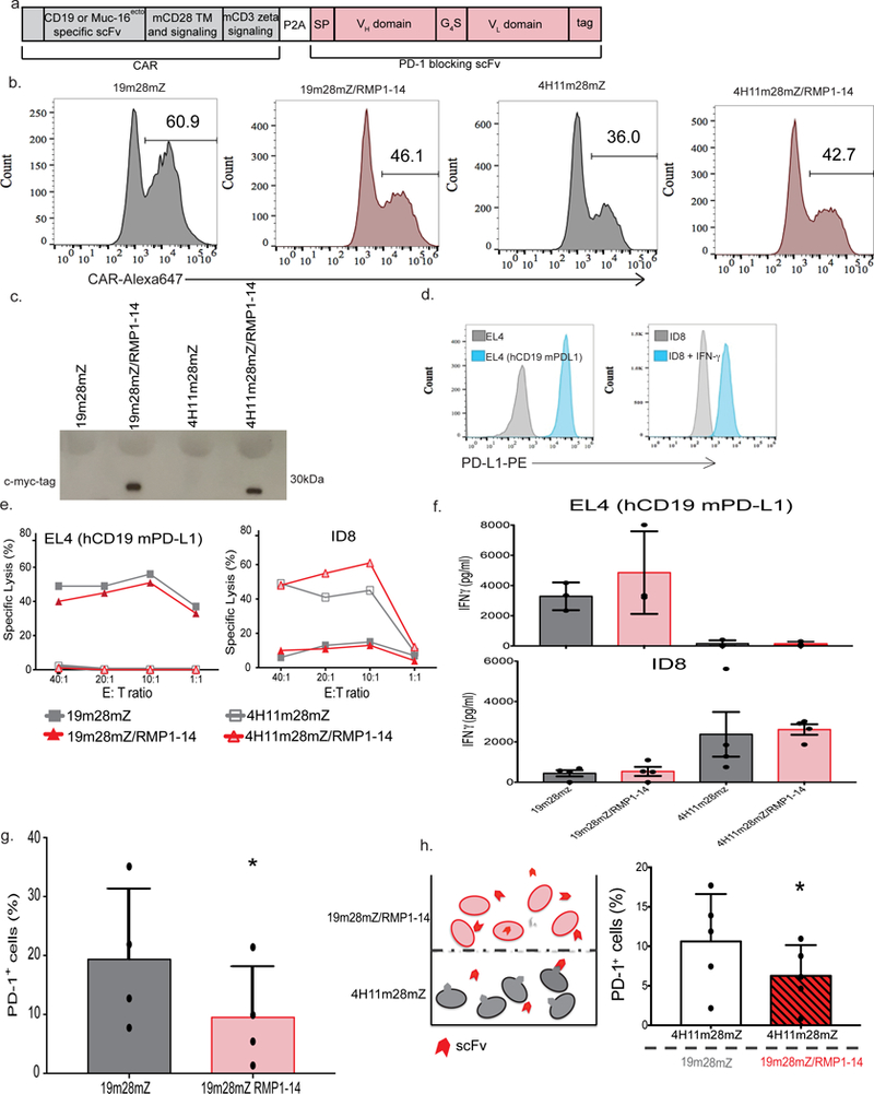
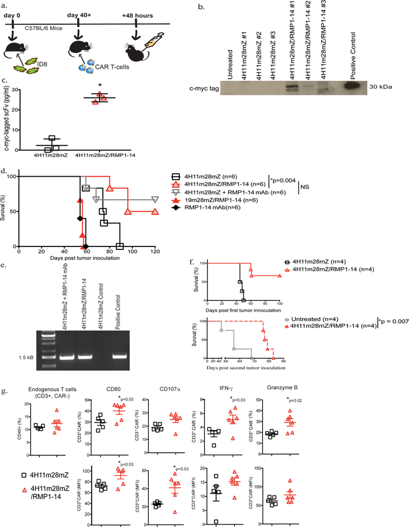
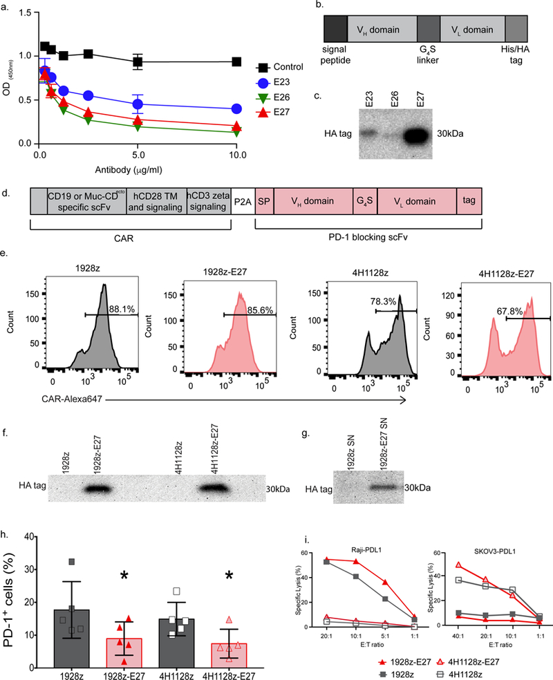
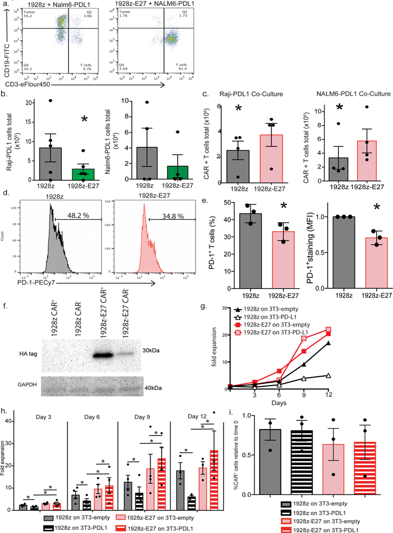
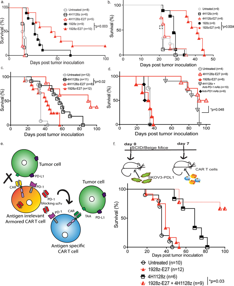
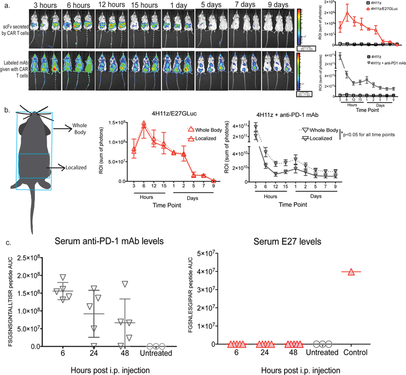
The efficacy of chimeric antigen receptor (CAR) T cell therapy against poorly responding tumors can be enhanced by administering the cells in combination with immune checkpoint blockade inhibitors. Alternatively, the CAR construct has been engineered to coexpress factors that boost CAR-T cell function in the tumor microenvironment. We modified CAR-T cells to secrete PD-1-blocking single-chain variable fragments (scFv). These scFv-secreting CAR-T cells acted in both a paracrine and autocrine manner to improve the anti-tumor activity of CAR-T cells and bystander tumor-specific T cells in clinically relevant syngeneic and xenogeneic mouse models of PD-L1+ hematologic and solid tumors. The efficacy was similar to or better than that achieved by combination therapy with CAR-T cells and a checkpoint inhibitor. This approach may improve safety, as the secreted scFvs remained localized to the tumor, protecting CAR-T cells from PD-1 inhibition, which could potentially avoid toxicities associated with systemic checkpoint inhibition.


Conflict of interest statement

Competing Financial Interests Statement: R.J.B. is a co-founder and receives royalties from Juno Therapeutics. R.J.B, H.J.J., and C.L. have submitted a patent related to this work.

Figures

Fig. 1.
Fig. 1.. Mouse CAR-T cells can be co-modified to secrete mouse PD-1-blocking scFv RMP1–14.
(a) Schematic of the bi-cistronic vectors utilized for syngeneic mouse studies encoding the CD19-targeted 19m28mZ CAR, or ovarian MUC16ecto-targeted 4H11m28mZ CAR, linked with a P2A element to the secretable PD-1-blocking scFv, RMP1–14. A c-myc-tag is included for detection of the scFv. (b) Representative flow cytometry plot demonstrating CAR expression following mouse T cell transduction, detected with fluorescently labeled CAR-specific idiotypic antibodies. Data shown is representative of ≥3 independent experiments. (c) Western blot on supernatant from equivalent numbers of viral packaging cells transduced to express the secretable scFv with the CAR, detected with anti-myc-tag antibody. Data shown is representative of 3 independent experiments. (d) Flow cytometry histograms depicting expression of mouse PD-L1 on EL4 (hCD19 mPD-L1) or ID8 cells. Data shown is representative of 3 independent experiments. (e) 4-hour 51Cr release assay demonstrating lysis of tumor cells. Data shown is representative of 3 independent experiments. (f) All 4 CAR constructs produce antigen-specific IFN-γ after co-culture with tumor cells. Data shown is mean +/− SEM from 4 independent experiments. (g) Quantification of PD-1 detection on CAR-T cells, as measured by flow cytometry. Data shown is mean +/− SEM from 4 independent experiments, *p=0.011 by two-tailed paired t-test. (h) Experimental schematic and quantification of decreased PD-1 detection by flow cytometry on 4H11m28mZ T cells when cultured in a transwell plate with 19m28mZ or 19m28mZ/RMP1–14 T cells. Data shown is mean +/− SEM from 5 separate donors, *p=0.012 by two-tailed paired t-test.
Fig. 2.
Fig. 2.. CAR-T cells secreting RMP1–14 scFv have enhanced anti-tumor function in syngeneic mouse tumor models.
(a) Schematic diagram of experimental setup to detect scFv secretion in vivo. C57BL/6 mice were inoculated with ID8 tumor, monitored until development of ascites and subsequently treated i.p. with 4H11m28mZ or 4H11m28mZ/RMP1–14 T cells. In vivo secretion of RMP1–14 scFv was detected by harvesting ascites from tumor-bearing mice 48 hours later. The ascites was immunoprecipitated with an anti-myc-tag antibody and (b) run on a Western blot using an anti-myc tag antibody or (c) run on Luminex utilizing anti-myc-tag beads (*p<0.0004 using an two-tailed unpaired t test, Mean ± SEM of 4H11m28mZ and 4H11m28mZ/RMP1–14 are 2.3 ± 1.9 and 26 ± 1.2, respectively), Data shown is from 2 independent experiments. (d) C57BL/6 mice were injected i.p. with ID8 tumor cells and treated with CAR-T cells, 250 μg RMP1–14 mAb or a combination of both 7 days later. RMP1–14 mAb was given on days 3, 7 and 14 post-tumor inoculation (*p=0.004 by Log-rank Mantel-Cox Test, with a 95% Confidence Interval (CI) of 0.4 to 0.9). Data shown is from 2 independent experiments. (e) PCR of bone marrow from mice surviving >120 days since tumor inoculation in Fig. 2d. CAR-T cells were detected in the bone marrow of long-term surviving mice treated with 4H11m28mZ T cells + RMP1–14 mAb or 4H11m28mZ/RMP1–14 T cells. Data shown is from 2 independent experiments. (f) C57BL/6 mice were inoculated with ID8 tumor and treated 7 days later with 4H11m28mZ or 4H11m28mZ/RMP1–14 T cells. Long term surviving mice in the 4H11m28mZ/RMP1–14 T cell cohort were re-challenged with a second inoculation of ID8 cells and compared to naïve untreated ID8-innoculated mice (*p=0.007 by Log-rank Mantel-Cox Test, with a 95% Confidence Interval of 0.2 to 0.3). (g) Quantification of flow cytometric analysis demonstrating endogenous, CAR- T cells extracted from C57BL/6 mice bearing B16-F10 mouse melanoma and treated with PD-1-blocking scFv CAR-T cells have enhanced activation and cytokines levels as compared to mice treated with second-generation CAR-T cells (*p values indicated on figure, by two-tailed unpaired t test). Data shown is pooled from 6 mice and 2 independent experiments.
Fig. 3.
Fig. 3.. Human CAR-T cells can be co-modified to secrete a novel PD-1-blocking scFv, E27.
(a) Novel human PD-1-blocking mAb candidates E27, E26 and E23 were utilized in a competitive binding assay to detect interruption of PD-1 binding to PD-L1 at varying concentrations, compared to a human IgG isotype control mAb (control). Data shown is mean of three independent experiments. (b) Schematic representation of PD-1-blocking scFv designed from the E23, E26 and E27 mAbs used in (c), where the signal peptide was linked to the variable heavy sequence, serine glycine linker and the variable light chain sequence. The His/HA tag was included for detection of the scFv. (c) Western blot on supernatant from equivalent numbers of 293-Glv9 packaging cells transduced to secrete scFvs with the 1928z CAR, stained with anti-HA antibody. Data shown is representative of 2 independent experiments. (d) Schematic of the bi-cistronic vector encoding the CD19-targeted 1928z CAR, or ovarian MUC16ecto-targeted 4H1128z CAR, linked with a P2A element to the secretable anti-human PD-1-blocking scFv, E27. (e) Representative flow cytometry plot demonstrating CAR expression following donor human T cell transduction, detected with fluorescently labeled CAR-specific idiotypic antibodies. Data shown is representative of ≥3 independent experiments. (f) Western blot on supernatant from CAR-T cells stained with anti-HA mAb, demonstrating a ~30 kDa protein in the 1928z-E27 and 4H1128z-E27 T cells. Data shown is representative of 2 independent experiments. (g) Western blot analysis of 293Glv9-PD-1+ cells incubated in supernatant from 1928z and 1928z-E27 T cells, stained with anti-HA mAb, showing a ~30 kDa protein in the PD-1+ cells incubated with supernatant from 1928z-E27. Data shown is representative of 2 independent experiments. (h) Quantification of PD-1 detection by flow cytometry on 1928z-E27 and 4H1128z-E27 T cell, as compared to second-generation CAR-T cells. Data shown is mean +/− SEM from 5 independent donors. For comparison of 1928z to 1928z-E27 *p=0.05, and 4h1128z to 4H1128z-E27 *p=0.006, both by a two-tailed paired t test. (i) 4-hour 51Cr release assay demonstrating that all 4 CAR constructs have antigen-dependent lysis of tumor cells. Data shown is representative of 3 independent donors and experiments.
Fig. 4.
Fig. 4.. Co-expression of CAR and E27 scFv protects proliferative and lytic capacity of T cells in the context of PD-L1+ tumor cells.
(a) Representative flow cytometry dot plots demonstrating lysis of Raji-PDL1 tumor cells, as determined by flow cytometry following 72 hour co-culture. Data shown is representative of 3 independent donors and experiments. (b). 1928z-E27 T cells lyse significantly more Raji-PDL1 tumor cells compared to 1928z T cells, data shown the mean +/− SEM from 5 independent experiments, *p=0.03 by a one-tailed paired t test. (c) CAR-T cells expansion numbers following co-culture with Raji-PDL1 or NALM6-PDL1 tumor cells as determined by flow cytometry, data shown is the average total number of T cells +/− SEM from 4 independent experiments, *p=0.05 for Raji experiment and *p=0.02 for Nalm6 experiment, both by a two-tailed paired t test. Representative flow cytometry plot (d) and quantification (e) showing increased PD-1 detection on 1928z T-cells compared to 1928z-E27 T-cells following 7 days co-culture with Raji-PDL1 tumor cells. Data shown in the mean +/− SEM from 3 independent experiments. *p=0.03 for percent positive CAR-T cell and MFI of staining, both by two-tailed paired test. (f) 1928z and 1928z-E27 T-cells were co-cultured with human T cells transduced to overexpress PD-1 and after 4 days stimulation with CD3/CD28 beads, the cells were sorted by flow cytometry to separate CAR+ and CAR- cells. Western blot was performed on the sorted populations and probed with anti-HA mAb. Data shown is representative of 3 independent donors and experiments. (g) Representative example of 1928z and 1928z-E27 T cells fold expansion when cultured with 3T3-empty or 3T3-PDL1 cells and stimulated with CD3/CD28 beads. Data shown is representative of 3 independent donors and experiments. (h) Cells were enumerated and re-plated on new 3T3 cells on days 3, 6, 9 and 12. 1928z T cells had reduced expansion when cultured with 3T3-PDL1 cells compared to 3T3- empty cells. 1928z-E27 cells had equivalent expansion when cultured on 3T3- empty or 3T3-PDL1 cells. Data shown is the mean fold expansion +/− SEM from 4 independent experiments, *p<0.05 by two-tailed paired t test. (i) Expansion of 1928z-E27 T cells on 3T3-PDL1 cells was due to an increase in both CAR+ and CAR- cells, comparing populations on day 0 and following expansion on 3T3-PDL1 cells at day 12. Data shown is representative of 3 independent experiments.
Fig. 5.
Fig. 5.. CAR-T cells that secrete E27 scFv have enhanced anti-tumor function in vivo.
SCID/Beige mice were inoculated with (a) Raji-PDL1 or (b) NALM6-PDL1 tumor cells and treated with 1928z-E27 CAR-T cells have enhanced survival over mice treated with 1928z (*p=0.003 with 95% CI of 0.1–0.9 and *p=0.0004 with 95% CI of 0.02–0.3, respectively by Log-rank Mantel-Cox test). Data shown is pooled from 2 independent experiments. (c) SCID/Beige mice treated with 4H1128z-E27 T cells had enhanced survival compared to mice treated with 4H1128z or irrelevant antigen 1928z-E27 T-cells in mice bearing SKOV3-PDL1 ovarian tumor cells. Data shown is from 2 independent experiments, *p=0.02 by Log-rank Mantel-Cox test with a 95% CI of 0.2–1.5. (d) Survival curve showing mice treated with 4H1128z-E27 T cells had enhanced survival compared to mice treated with 4H1128z + anti-human PD-1 mAb. Data shown is from 2 independent experiments, *p=0.05 by Log-rank Mantel-Cox test with a 95% CI of 0.4–3.1. (e) Schematic illustration of experiment to study bystander effect of scFv-secreting CAR-T cells where SKOV3-PDL1 tumor-bearing mice were treated with a combination of E27-secreting 1928z T-cells, which are antigen irrelevant in this model, together with ovarian-tumor specific 4H1128z T cells. PD-1-blocking scFv secreted by the 1928z-E27 T cells in vivo will bind to PD-1 on 4H1128z T-cells and enhance tumor-specific function. (f) Mice treated 7 days after SKOV3-PDL1 inoculation with a mix of 1928z-E27 + 4H1128z T-cells have enhanced survival as compared to mice treated with 4H1128z (*p=0.007, 95% CI of 0.03–0.6) or 1928z-E27 (*p<0.0001, 95% CI of 0.02–0.2) T cells alone. Data shown is from 2 independent experiments.
Fig. 6.
Fig. 6.. The PD-1-blocking E27 scFv secreted by CAR-T cells is only detected in the local tumor microenvironment.
(a) Imaging and quantification over time of E27 scFv tagged with Gaussia Luciferase (GLuc) or fluorescently labeled anti-human PD-1 mAb in SKOV3-PDL1 tumor-bearing SCID/Beige mice treated i.p. with scFv-secreting CAR-T cells or CAR-T cells with anti-PD-1 mAb, 3 animals per group. (b) Schematic representation and quantitation of areas utilized for complete versus local detection of scFv or antibody. *p<0.02 at all time points tested for antibody using multiple T test with 3 animals per group. (c) Quantification utilizing unique peptide sequences by liquid chromatography-tandem mass spectrometry (LC-MS/MS) of serum levels over time of E27 scFv and anti-human PD-1 mAb in in SKOV3-PDL1 tumor-bearing SCID/Beige mice treated i.p. with scFv-secreting CAR-T cells or CAR-T cells with anti-PD-1 mAb (5 animals per group). Systemic infusion of isolated E27 scFv was utilized as a positive control.

References

    1. Jackson HJ, Rafiq S & Brentjens RJ Driving CAR-T cells forward. Nature reviews. Clinical oncology 13, 370–383 (2016). - PMC - PubMed
    1. Zhang M, et al. A high M1/M2 ratio of tumor-associated macrophages is associated with extended survival in ovarian cancer patients. J Ovarian Res. 7:19 (2014). - PMC - PubMed
    1. Knutson KL, et al. Regulatory T cells, inherited variation, and clinical outcome in epithelial ovarian cancer. Cancer Immunol Immunother 12, 1495–504 (2015). - PMC - PubMed
    1. Erfani N, et al. FoxP3+ regulatory T cells in peripheral blood of patients with epithelial ovarian cancer. Iran J Immunol 2, 105–12 (2014). - PubMed
    1. Shevach EM Mechanisms of foxp3+ T regulatory cell-mediated suppression. Immunity 30(5), 636–45 (2009). - PubMed

Methods-only References

    1. Peterson AC, Russell JD, Bailey DJ, Westphall MS & Coon JJ Parallel reaction monitoring for high resolution and high mass accuracy quantitative, targeted proteomics. Mol Cell Proteomics 11, 1475–1488 (2012). - PMC - PubMed
    1. Egertson JD, MacLean B, Johnson R, Xuan Y & MacCoss MJ Multiplexed peptide analysis using data-independent acquisition and Skyline. Nat Protoc 10, 887–903 (2015). - PMC - PubMed
    1. Shevchenko A, Tomas H, Havlis J, Olsen JV & Mann M In-gel digestion for mass spectrometric characterization of proteins and proteomes. Nat Protoc 1, 2856–2860 (2006). - PubMed
    1. Rappsilber J, Mann M & Ishihama Y Protocol for micro-purification, enrichment, pre-fractionation and storage of peptides for proteomics using StageTips. Nat Protoc 2, 1896–1906 (2007). - PubMed
    1. Lee AY et al. Measurement of fractional synthetic rates of multiple protein analytes by triple quadrupole mass spectrometry. Clin Chem 58, 619–627 (2012). - PubMed

Publication types

Substances