

Longitudinal 18F-FDG PET imaging in a rat model of autoimmune myocarditis
- PMID: 30102319
- PMCID: PMC6429237
- DOI: 10.1093/ehjci/jey119
Longitudinal 18F-FDG PET imaging in a rat model of autoimmune myocarditis
Abstract
Aims: Although mortality rate is very high, diagnosis of acute myocarditis remains challenging with conventional tests. We aimed to elucidate the potential role of longitudinal 2-Deoxy-2-18F-fluoro-D-glucose (18F-FDG) positron emission tomography (PET) inflammation monitoring in a rat model of experimental autoimmune myocarditis.
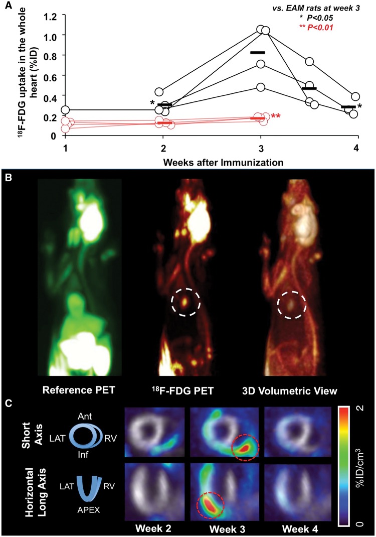
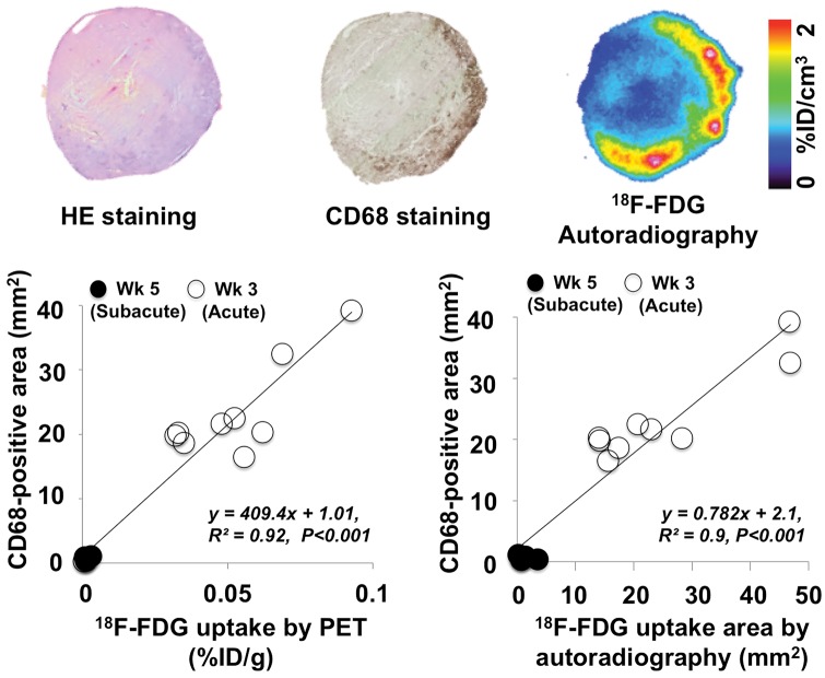
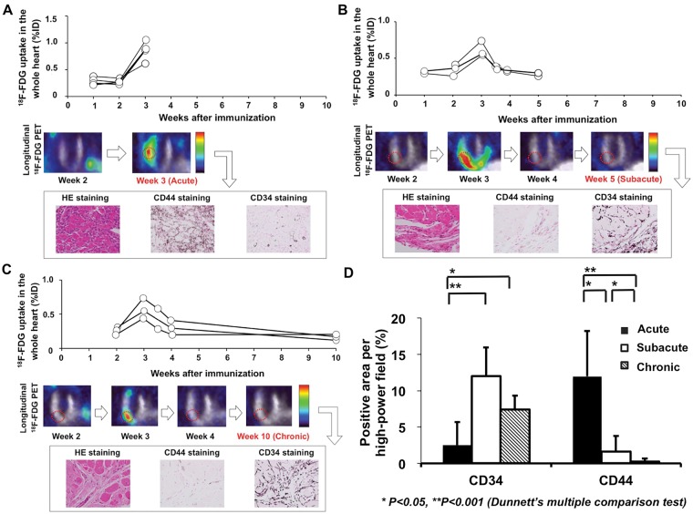
Methods and results: Autoimmune myocarditis was induced in Lewis rats by immunizing with porcine cardiac myosin emulsified in complete Freund's adjuvant. Time course of disease was assessed by longitudinal 18F-FDG PET imaging. A correlative analysis between in- and ex vivo18F-FDG signalling and macrophage infiltration using CD68 staining was conducted. Finally, immunohistochemistry analysis of the cell-adhesion markers CD34 and CD44 was performed at different disease stages determined by longitudinal 18F-FDG PET imaging. After immunization, myocarditis rats revealed a temporal increase in 18F-FDG uptake (peaked at week 3), which was followed by a rapid decline thereafter. Localization of CD68 positive cells was well correlated with in vivo18F-FDG PET signalling (R2 = 0.92) as well as with ex vivo18F-FDG autoradiography (R2 = 0.9, P < 0.001, respectively). CD44 positivity was primarily observed at tissue samples obtained at acute phase (i.e. at peak 18F-FDG uptake), while CD34-positive staining areas were predominantly identified in samples harvested at both sub-acute and chronic phases (i.e. at 18F-FDG decrease).
Conclusion: 18F-FDG PET imaging can provide non-invasive serial monitoring of cardiac inflammation in a rat model of acute myocarditis.
Keywords: 18F-FDG; PET; inflammation; myocarditis; personalized treatment.
© The Author(s) 2018. Published by Oxford University Press on behalf of the European Society of Cardiology.
Figures
References
-
- Magnani JW, Dec GW.. Myocarditis: current trends in diagnosis and treatment. Circulation 2006;113:876–90. - PubMed
-
- Noren GR, Staley NA, Bandt CM, Kaplan EL.. Occurrence of myocarditis in sudden death in children. J Forensic Sci 1977;22:188–96. - PubMed
-
- Frick M, Pachinger O, Polzl G.. [Myocarditis and sudden cardiac death in athletes. Diagnosis, treatment, and prevention]. Herz 2009;34:299–304. - PubMed
-
- Mahrholdt H, Sechtem U.. Noninvasive differentiation between active and healed myocarditis by cardiac magnetic resonance: are we there yet? JACC Cardiovascular Imaging 2009;2:139–42. - PubMed
-
- Skouri HN, Dec GW, Friedrich MG, Cooper LT.. Noninvasive imaging in myocarditis. J Am Coll Cardiol 2006;48:2085–93. - PubMed
MeSH terms
Substances
LinkOut - more resources
Full Text Sources
Other Literature Sources
Miscellaneous
