. 2018 Oct 12;46(18):9816-9828.
doi: 10.1093/nar/gky723.

p15PAF binding to PCNA modulates the DNA sliding surface

Affiliations

p15PAF binding to PCNA modulates the DNA sliding surface

Matteo De March et al. Nucleic Acids Res. .

Abstract

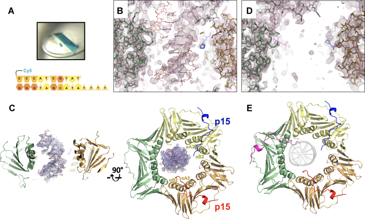
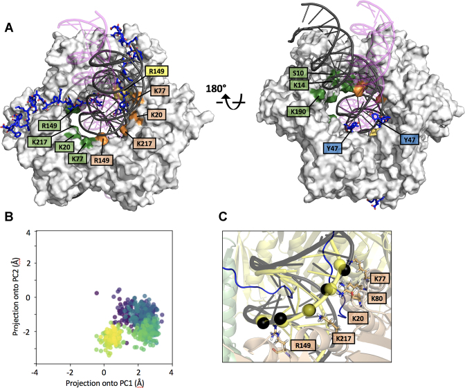
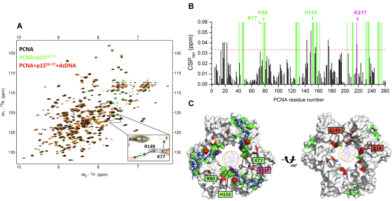
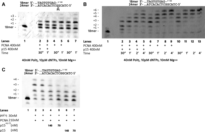
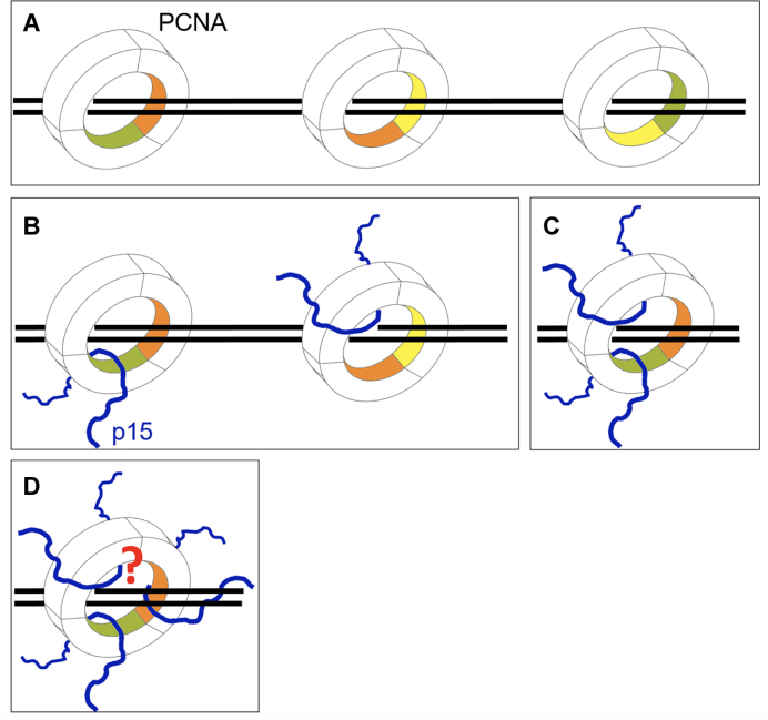
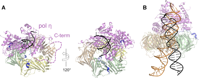
p15PAF is an oncogenic intrinsically disordered protein that regulates DNA replication and lesion bypass by interacting with the human sliding clamp PCNA. In the absence of DNA, p15PAF traverses the PCNA ring via an extended PIP-box that contacts the sliding surface. Here, we probed the atomic-scale structure of p15PAF-PCNA-DNA ternary complexes. Crystallography and MD simulations show that, when p15PAF occupies two subunits of the PCNA homotrimer, DNA within the ring channel binds the unoccupied subunit. The structure of PCNA-bound p15PAF in the absence and presence of DNA is invariant, and solution NMR confirms that DNA does not displace p15PAF from the ring wall. Thus, p15PAF reduces the available sliding surfaces of PCNA, and may function as a belt that fastens the DNA to the clamp during synthesis by the replicative polymerase (pol δ). This constraint, however, may need to be released for efficient DNA lesion bypass by the translesion synthesis polymerase (pol η). Accordingly, our biochemical data show that p15PAF impairs primer synthesis by pol η-PCNA holoenzyme against both damaged and normal DNA templates. In light of our findings, we discuss the possible mechanistic roles of p15PAF in DNA replication and suppression of DNA lesion bypass.


Figures

Figure 1.
Figure 1.
Crystal structures of human PCNA bound to p15 fragments and DNA. (A) Blue crystals of p1550–77–PCNA–DNA complex. Co-crystals of PCNA mixed with p1541–72 and DNA were also blue, confirming incorporation of DNA in the crystal lattice. The cartoon below shows the sequence of the DNA substrate. (B) Side view of the 2Fo – Fc omit map of the p1550–77–PCNA–DNA complex refined without DNA in the model, contoured at 0.7 σ, showing the PCNA central channel. PCNA subunits (green and wheat) and p1550–77 peptide in the background (blue) are in stick representation. The loop of a symmetry related PCNA molecule is shown in grey. The DNA, modeled as in the PCNA–dsDNA binary structure (5), is shown in orange. (C) Side- and top views of the refined p1550–77–PCNA–DNA complex structure. PCNA and p1550–77 are shown in ribbon representation, and the protein and peptide chains colored differently. The DNA, shown in orange, is modeled as in the PCNA–dsDNA binary structure. The 2Fo – Fc map around DNA is shown contoured at 0.7σ. (D) Side view of the 2Fo – Fc map of the p1541–72–PCNA complex contoured at 0.7σ, showing the PCNA central channel as in (B). (E) Top view of p1541–72–PCNA complex structure, color-coded as in (C). The DNA shown as a grey transparent ribbon in the same position as in (C) would cause a steric clash with the N-terminus of the p15 peptide on the third PCNA subunit.
Figure 2.
Figure 2.
MD simulation of PCNA bound to two p1547–70 peptides and a 40 bp DNA (A) Superposition of the initial and equilibrium states of the MD trajectory. PCNA is shown as a gray surface and DNA as a ribbon. The DNA in magenta (with transparency), and black correspond to the initial and equilibrium states of the simulation, respectively. PCNA residues whose side chains are engaged in polar contacts with DNA phosphates are labeled. Residues of different PCNA subunits are colored in green, yellow and wheat. (B) Principal Component Analysis of the evolution of the DNA position inside the PCNA ring (see Methods section for details). The centre of DNA in each trajectory frame was projected onto the first 2 components of the subspace composed of the centres of the 3 PCNA subunits. Each frame is coloured using the viridis colormap, which goes from dark purple for the first frames to yellow for the last ones. In the initial frame, DNA is close to (0,0), the centre of the three PCNA chains, and it quickly translates to a non-centered position. The final position is retained due to the stabilizing interactions reported in Supplementary Figure S4. (C) Close-up of the equilibrium state of the MD trajectory showing the PCNA–DNA interface. Interacting PCNA side chains and DNA phosphates (interatomic side chain nitrogen – DNA phosphorus distance < 4 Å) are shown as sticks and black spheres, respectively. DNA in yellow corresponds to the position in the crystallographic PCNA–dsDNA binary structure (5), with interfacial phosphates shown as spheres.
Figure 3.
Figure 3.
NMR analysis of PCNA binding to p1550–77 and a 10 bp dsDNA. (A) Superposition of 1H-15N TROSY spectra of 95 μM PCNA in the absence (black) and presence (green) of 606 μM of p1550–77 and (red) of 92 μM dsDNA (left) generated with oligonucleotides 3–4 in Supplementary Table S1. Spectra were acquired at 35°C on samples in 20 mM sodium phosphate, 50 mM NaCl, pH 7.0. The expansion shows signals of three representative residues. A96 signal is not perturbed by the addition of either p1550–77 or DNA. R149 signal persists upon p1550–77 addition, and shifts significantly by the sequential addition of DNA. Conversely, K77 signal disappears at substoichiometric concentrations of p1550–77, and is not recovered by DNA addition. The dotted arrow points to a signal that is tentatively assigned to K77 in the p1550–77-bound form. (B) Chemical shift perturbations (CSP) of backbone amide 1H and 15N NMR resonances induced by DNA. The dotted line indicates the average plus two standard deviations. The green bars indicate the position of residues that disappear upon addition of substoichiometric p1550–77, and are not drawn to scale. The residues perturbed by p1550–77 and that also appear at the interface of the p1550–77–PCNA–DNA crystal structure are labeled. (C) Front- and back-face views of PCNA surface. PCNA residues whose amide signals disappear in the presence of substoichiometric p1550–77, or are significantly perturbed by DNA are colored green or red, respectively. p1550–77 at the three PCNA PIP-box sites is shown in sticks, and DNA in the crystallographic position is shown as and orange ribbon.
Figure 4.
Figure 4.
Inhibition of pol η holoenzyme by p15 (A) Time course of the reaction of pol η in the presence of PCNA/p15 at equimolar concentrations (Lanes 2 and 3), in the presence of PCNA (Lanes 4 and 5), or with pol η alone (lanes 6 and 7) on a cisPt(GG) template (10 nM), with all four dNTPs at the indicated concentration. (B) Time course of the reaction of pol η on the template without the lesion (10 nM), in the presence of PCNA/p15 at equimolar concentrations (lanes 2–5), in the presence of PCNA (lanes 6–9), or pol η alone (lanes 10–13), with all four dNTPs at the indicated concentration. (C) Reaction of pol η replicating the undamaged template in the presence of PCNA and in the absence or presence of p1541–72 peptide or full length p15. Reactants at the indicated concentrations were incubated at 37°C for 30 s and the reaction was stopped by addition of standard denaturing gel loading buffer. In all these experiments, PCNA was not ubiquitylated. These experiments show that p15 downregulates the activity of pol η–PCNA holoenzyme in bypassing a cisplatin lesion as well as in replicating a normal DNA substrate.
Figure 5.
Figure 5.
Possible effects of p15 on PCNA sliding (A) PCNA can diffuse on DNA contacting three equivalent sliding surfaces, each composed of two homologous sets of basic residues spanning across the interface of two subunits (the 3 PCNA subunits are colored green, yellow and wheat). (B–D) The stoichiometry of p15 binding to the PCNA homotrimer defines the available surfaces for clamp sliding. Whether a configuration where PCNA simultaneously binds three p15 chains and DNA, can be achieved, and whether it completely or partially hinders sliding, remains to be determined.
Figure 6.
Figure 6.
Structural models of pol η-PCNA holoenzymes with p15 and DNA. (A) The PCNA trimer of the p1550–77–PCNA crystal structure (PDB ID: 4D2G) was superposed to PCNA of the low-resolution structure of human pol η–PCNA–DNA generated from EM data (PDB ID: 3JA9 and 3JAA) (33). DNA is shown in black. The vacant PIP-box site on PCNA (subunit wheat) was occupied by the C-terminal PIP-box of pol η using the crystal structure of human PCNA bound to pol η residues 700–710 (chain W of PDB ID 2ZVK) (40). The dashed line indicates the flexible pol η C-terminus (residues 433–699). (B) The PCNA trimer of the structure of p1550–77–PCNA–DNA complex (PDB: 6EHT) was superposed to PCNA of the pol η–PCNA–DNA (48) complex (PDB: 3JA9 and and 3JAA). The DNA of the first complex (elongated to 40 bp) is shown as an orange ribbon, that of the latter (elongated to 25 bp) as a black ribbon. According to these models, it is possible that p15 may co-exist with pol η on the same PCNA ring. However, the constraint on DNA within the clamp channel imposed by p15 may hinder the translocation of pol η holoenzyme on DNA.

References

    1. Krishna T.S., Kong X.P., Gary S., Burgers P.M., Kuriyan J.. Crystal structure of the eukaryotic DNA polymerase processivity factor PCNA. Cell. 1994; 79:1233–1243. - PubMed
    1. McNally R., Bowman G.D., Goedken E.R., O’Donnell M., Kuriyan J.. Analysis of the role of PCNA-DNA contacts during clamp loading. BMC Struct. Biol. 2010; 10:3. - PMC - PubMed
    1. Mailand N., Gibbs-Seymour I., Bekker-Jensen S.. Regulation of PCNA-protein interactions for genome stability. Nat. Rev. Mol. Cell Biol. 2013; 14:269–282. - PubMed
    1. De Biasio A., de Opakua A.I., Mortuza G.B., Molina R., Cordeiro T.N., Castillo F., Villate M., Merino N., Delgado S., Gil-Carton D. et al. . Structure of p15(PAF)-PCNA complex and implications for clamp sliding during DNA replication and repair. Nat. Commun. 2015; 6:6439. - PubMed
    1. De March M., Merino N., Barrera-Vilarmau S., Crehuet R., Onesti S., Blanco F.J., De Biasio A.. Structural basis of human PCNA sliding on DNA. Nat. Commun. 2017; 8:13935. - PMC - PubMed

Publication types

MeSH terms

LinkOut - more resources