Uteroplacental and Fetal 4D Flow MRI in the Pregnant Rhesus Macaque
- PMID: 30102431
- PMCID: PMC6330144
- DOI: 10.1002/jmri.26206
Uteroplacental and Fetal 4D Flow MRI in the Pregnant Rhesus Macaque
Abstract
Background: Pregnancy complications are often associated with poor uteroplacental vascular adaptation and standard diagnostics are unable to reliably quantify flow in all uteroplacental vessels and have poor sensitivity early in gestation.
Purpose: To investigate the feasibility of using 4D flow MRI to assess total uteroplacental blood flow in pregnant rhesus macaques as a precursor to human studies.
Study type: Retrospective feasibility study.
Animal model: Fifteen healthy, pregnant rhesus macaques ranging from the 1st trimester to 3rd trimester of gestation.
Field strength/sequence: Abdominal 4D flow MRI was performed on a 3.0T scanner with a radially undersampled phase contrast (PC) sequence. Reference ferumoxytol-enhanced angiograms were acquired with a 3D ultrashort echo time sequence with a center-out radial trajectory.
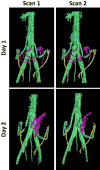
Assessment: Repeatability of flow measurements was assessed with scans performed same-day and on consecutive days in the uterine arteries and ovarian veins. In-flow was compared against out-flow in the uterus, umbilical cord, and fetal heart with a conservation of mass analysis. Conspicuity of uteroplacental vessels was qualitatively compared between PC angiograms derived from 4D flow data and ferumoxytol-enhanced angiograms.
Statistical tests: Bland-Altman analysis was used to quantify same-day and consecutive-day repeatability.
Results: Same-day flow measurements showed an average difference between scans of 13% in both the uterine arteries and ovarian veins, while consecutive-day measurements showed average differences of 22% and 24%, respectively. Comparisons of in-flow and out-flow showed average differences of 15% in the uterus, 8% in fetal heart, and 15% in the umbilical cord. PC angiograms showed similar depiction of main uteroplacental vessels as high-resolution, ferumoxytol-enhanced angiograms.
Data conclusion: 4D flow MRI could be used in the rhesus macaque for repeatable flow measurements in the uteroplacental and fetal vasculature, setting the stage for future human studies.
Level of evidence: 2 Technical Efficacy: Stage 1 J. Magn. Reson. Imaging 2019;49:534-545.
© 2018 International Society for Magnetic Resonance in Medicine.
Figures


Similar articles
-
Perfusion of the placenta assessed using arterial spin labeling and ferumoxytol dynamic contrast enhanced magnetic resonance imaging in the rhesus macaque.Magn Reson Med. 2019 Mar;81(3):1964-1978. doi: 10.1002/mrm.27548. Epub 2018 Oct 25. Magn Reson Med. 2019. PMID: 30357902 Free PMC article.
-
Evaluation of a motion-robust 2D chemical shift-encoded technique for R2* and field map quantification in ferumoxytol-enhanced MRI of the placenta in pregnant rhesus macaques.J Magn Reson Imaging. 2020 Feb;51(2):580-592. doi: 10.1002/jmri.26849. Epub 2019 Jul 5. J Magn Reson Imaging. 2020. PMID: 31276263 Free PMC article.
-
An MRI approach to assess placental function in healthy humans and sheep.J Physiol. 2021 May;599(10):2573-2602. doi: 10.1113/JP281002. Epub 2021 Mar 29. J Physiol. 2021. PMID: 33675040
-
4D flow MRI.J Magn Reson Imaging. 2012 Nov;36(5):1015-36. doi: 10.1002/jmri.23632. J Magn Reson Imaging. 2012. PMID: 23090914 Review.
-
Computational modeling of the interactions between the maternal and fetal circulations in human pregnancy.WIREs Mech Dis. 2021 Jan;13(1):e1502. doi: 10.1002/wsbm.1502. Epub 2020 Aug 3. WIREs Mech Dis. 2021. PMID: 32744412 Review.
Cited by
-
Impact of ferumoxytol magnetic resonance imaging on the rhesus macaque maternal-fetal interface†.Biol Reprod. 2020 Feb 14;102(2):434-444. doi: 10.1093/biolre/ioz181. Biol Reprod. 2020. PMID: 31511859 Free PMC article.
-
Fetal whole heart blood flow imaging using 4D cine MRI.Nat Commun. 2020 Oct 5;11(1):4992. doi: 10.1038/s41467-020-18790-1. Nat Commun. 2020. PMID: 33020487 Free PMC article.
-
Nonhuman Primates in Translational Research.Annu Rev Anim Biosci. 2022 Feb 15;10:441-468. doi: 10.1146/annurev-animal-021419-083813. Annu Rev Anim Biosci. 2022. PMID: 35167321 Free PMC article. Review.
-
The application of in utero magnetic resonance imaging in the study of the metabolic and cardiovascular consequences of the developmental origins of health and disease.J Dev Orig Health Dis. 2021 Apr;12(2):193-202. doi: 10.1017/S2040174420001154. Epub 2020 Dec 14. J Dev Orig Health Dis. 2021. PMID: 33308364 Free PMC article. Review.
-
The current state and potential innovation of fetal cardiac MRI.Front Pediatr. 2023 Jul 14;11:1219091. doi: 10.3389/fped.2023.1219091. eCollection 2023. Front Pediatr. 2023. PMID: 37520049 Free PMC article. Review.
References
Publication types
MeSH terms
Substances
Grants and funding
- R01 HL117341/HL/NHLBI NIH HHS/United States
- UL1 TR000427/TR/NCATS NIH HHS/United States
- P51OD011106 (to the Wisconsin National Primate Center)/National Institutes of Health (NIH)/International
- U01 HD087216/HD/NICHD NIH HHS/United States
- NIH CTSA UW-ICTR UL1TR000427/National Institutes of Health (NIH)/International
LinkOut - more resources
Full Text Sources
Other Literature Sources
Medical
Research Materials
Miscellaneous

