. 2018 Nov;22(11):5429-5438.
doi: 10.1111/jcmm.13814. Epub 2018 Aug 13.

BMP-2 induces human mononuclear cell chemotaxis and adhesion and modulates monocyte-to-macrophage differentiation

Affiliations

BMP-2 induces human mononuclear cell chemotaxis and adhesion and modulates monocyte-to-macrophage differentiation

Evangelia Pardali et al. J Cell Mol Med. 2018 Nov.

Abstract

Type 2 diabetes mellitus (T2DM) is a cardiovascular risk factor which leads to atherosclerosis, an inflammatory disease characterized by the infiltration of mononuclear cells in the vessel. Bone morphogenetic protein (BMP)-2 is a cytokine which has been recently shown to be elevated in atherosclerosis and T2DM and to contribute to vascular inflammation. However, the role of BMP-2 in the regulation of mononuclear cell function remains to be established. Herein, we demonstrate that BMP-2 induced human monocyte chemotaxis via phosphoinositide 3 kinase and mitogen-activated protein kinases. Inhibition of endogenous BMP-2 signalling, by Noggin or a BMP receptor inhibitor, interfered with monocyte migration. Although BMP-2 expression was increased in monocytes from T2DM patients, it could still stimulate their migration. Furthermore, BMP-2 interfered with their differentiation into M2 macrophages. Finally, BMP-2 both induced the adhesion of monocytes to fibronectin and endothelial cells (ECs), and promoted the adhesive properties of ECs, by increasing expression of adhesion and pro-inflammatory molecules. Our data demonstrate that BMP-2 could exert its pro-inflammatory effects by inducing monocyte migration and adhesiveness to ECs and by interfering with the monocyte differentiation into M2 macrophages. Our findings provide novel insights into the mechanisms by which BMP-2 may contribute to the development of atherosclerosis.

Keywords: BMP; adhesion; atherosclerosis; chemotaxis; diabetes; endothelial cells; monocytes.


Figures

Figure 1
Figure 1
BMP‐2 induces monocyte migratory responses in a dose‐dependent manner via induction of PI3K, p38 and ERK signalling cascades. A, Monocytes were characterized for their chemotactic activity towards different concentrations of BMP‐2 and MCP‐1 (10 ng/mL). Noggin, a BMP‐2/4/7 inhibitor, inhibited the effect of BMP‐2 on monocyte migration (n = 5). B, Monocytes were treated with 500 ng/mL noggin or with 3 μmol/L LDN193189 for 30 min, and they were characterized for their basal and MCP‐1‐induced (10 ng/mL) chemotactic activity. (n = 5). C, Monocytes were stimulated with 100 ng/mL BMP‐2 for 10 min. Phosphorylation of Smad1, p38, ERK1/2 and AKT was determined by Western blot. D, Monocytes were incubated for 30 min with vehicle or different PI3K kinase inhibitors: 25 nmol/L of Wortmannin or 1 μmol/L LY29400 and they were assessed for their migratory responses towards 100 ng/mL BMP‐2 (n = 5). E, Monocytes were incubated for 30 min with vehicle or different MAPK kinase inhibitors, 10 μmol/L SB239063, 25 μmol/L PD98059 or 10 μmol/L U0126 and characterized for their migratory responses towards 100 ng/mL BMP‐2 (n = 5). Data are presented as mean ± SEM (**P < 0.01, ***P < 0.001)
Figure 2
Figure 2
T2DM interferes with VEGFA‐induced, but not with BMP‐2‐induced monocyte migratory responses and enhances BMP‐2 gene expression in monocytes. A. Monocytes were isolated from the blood of patients with T2DM (n = 10) or without T2DM (nT2DM, n = 20), and the mRNA expression of BMP‐2 was analysed by qRTPCR. B, C. Monocytes were isolated from patients with T2DM (n = 12) or without T2DM (nT2DM, n = 20). The monocyte chemotactic responses towards BMP‐2 (B) and VEGFA (C) were analysed. Data are represented as mean ± SEM. (**P < 0.01, ***P < 0.001)
Figure 3
Figure 3
BMP‐2 enhances the expression of macrophage markers, but interferes with the expression of M2 macrophages markers. A, B, Monocyte differentiation into macrophages was induced by addition of 50 ng/mL M‐CSF in the presence or absence of BMP‐2. Differentiation into M1 and M2 macrophages was induced with 10 ng/mL IL‐1β or 10 ng/ml IL‐4, respectively, in the presence or absence of BMP‐2. A, Morphology of monocyte‐derived M0, M1 and M2 macrophages was revealed by phase contrast microscopy. B‐C, The expression of the CD36 macrophage marker, M1 markers Il‐1β and M2 macrophage markers AMAC1 and CD163 was analysed by qRTPCR (n = 5). Data are represented as mean ± SEM. (**P < 0.01, ***P < 0.001)
Figure 4
Figure 4
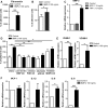
BMP‐2 induces mononuclear cell adhesiveness in human endothelial cells (ECs). A, Monocytes were incubated for 30 min with or without 100 ng/mL BMP‐2 and allowed to adhere on Fibronectin‐coated plates for 30 min, and the number of adherent cells was counted (n = 5). B, HUVECs were stimulated with 10 ng/mL Tumour necrosis factor‐α (TNF‐α) or 10 ng/mL MCP‐1 for 5 h. The mRNA expression of BMP‐2 was analysed by qRTPCR (n = 5). C, Freshly isolated monocytes were stimulated for 30 min with 100 ng/mL BMP‐2, labelled with Calcein and allowed to adhere on HUVECs, adherent cells were quantified (n = 5). D, HUVECs were preincubated with vehicle, 3 μmol/L LDN193189 BMP receptor kinase inhibitor, 1 μmol/L LY29400 PI3K inhibitor, 10 μmol/L SB239063 p38 kinase inhibitor or 25 μmol/L PD98059 MAPK inhibitor and then stimulated with 100 ng/mL BMP‐2 for 5 h. Labelled monocytes were allowed to adhere on them for 20 min, and adherent cells were quantified (n = 5). E‐F, HUVECs were stimulated with 100 ng/mL BMP‐2 for 5 h. The mRNA expression of ICAM‐1, VCAM‐1, MCP‐1, IL‐1β, IL‐6 and IL‐8 was analysed by qRTPCR (n = 6). Data are represented as mean ± SEM. (*P < 0.05, **P < 0.01, ***P < 0.001)
Figure 5
Figure 5
BMP‐2 induces endothelial cell adhesiveness in mouse endothelial cells (ECs). A, bEnd5 cells were preincubated with vehicle, 3 μmol/L LDN193189 BMP receptor kinase inhibitor, 1 μmol/L LY29400 PI3K inhibitor, 10 μmol/L SB239063 p38 kinase inhibitor or 25 μmol/L PD98059 MAPK inhibitor and then stimulated with 100 ng/mL BMP‐2. Calcein‐labelled primary monocytes were allowed to adhere on them for 20 min. Nonadherent cells were washed, and adherent cells were quantified using ImageJ (n = 5). B, bEnd5 cells were stimulated with 100 ng/mL BMP‐2 for 20 min. Protein lysates were analysed by Western blot for phosphorylation of downstream signalling regulators. Smad1, p38, ERK1/2. C, bEnd5 cells were stimulated with 100 ng/mL BMP‐2 for 5 h. The mRNA expression of ICAM‐1, VCAM‐1, MCP‐1 and IL‐6 was analysed by qRTPCR (n = 6). Data are represented as mean ± SEM. (*P < 0.05, **P < 0.01, ***P < 0.001)

References

    1. Libby P. Inflammation in atherosclerosis. Arterioscler Thromb Vasc Biol. 2012;32:2045‐2051. - PMC - PubMed
    1. Vattikuti R, Towler DA. Osteogenic regulation of vascular calcification: an early perspective. Am J Physiol Endocrinol Metab. 2004;286:E686‐E696. - PubMed
    1. Pardali E, Waltenberger J. Monocyte function and trafficking in cardiovascular disease. Thromb Haemost. 2012;108:804‐811. - PubMed
    1. Tchaikovski V, Olieslagers S, Bohmer FD, Waltenberger J. Diabetes mellitus activates signal transduction pathways resulting in vascular endothelial growth factor resistance of human monocytes. Circulation. 2009;120:150‐159. - PubMed
    1. Cai J, Pardali E, Sanchez‐Duffhues G, ten Dijke P. Bmp signaling in vascular diseases. FEBS Lett. 2012;586:1993‐2002. - PubMed

Publication types

MeSH terms