. 2018 Aug 13;14(8):e1007244.
doi: 10.1371/journal.ppat.1007244. eCollection 2018 Aug.

Macrophage-derived LTB4 promotes abscess formation and clearance of Staphylococcus aureus skin infection in mice

Affiliations

Macrophage-derived LTB4 promotes abscess formation and clearance of Staphylococcus aureus skin infection in mice

Stephanie L Brandt et al. PLoS Pathog. .

Abstract

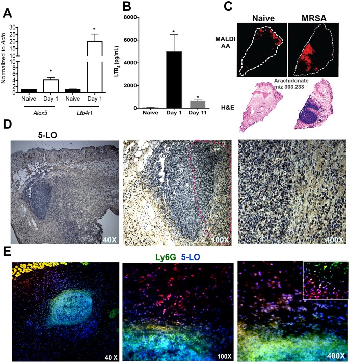
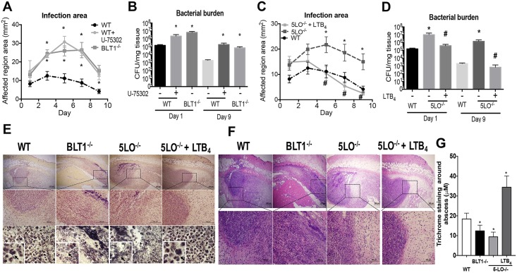
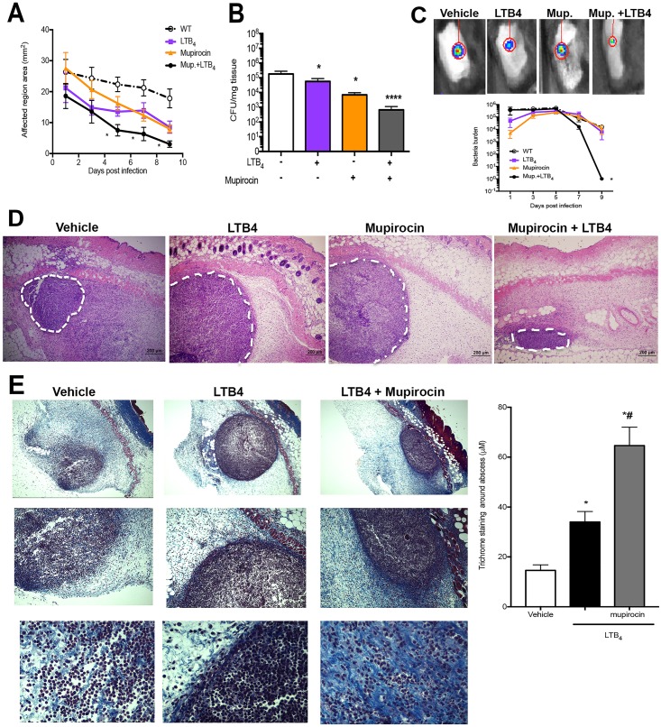
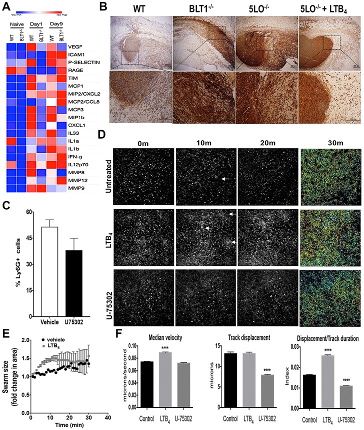
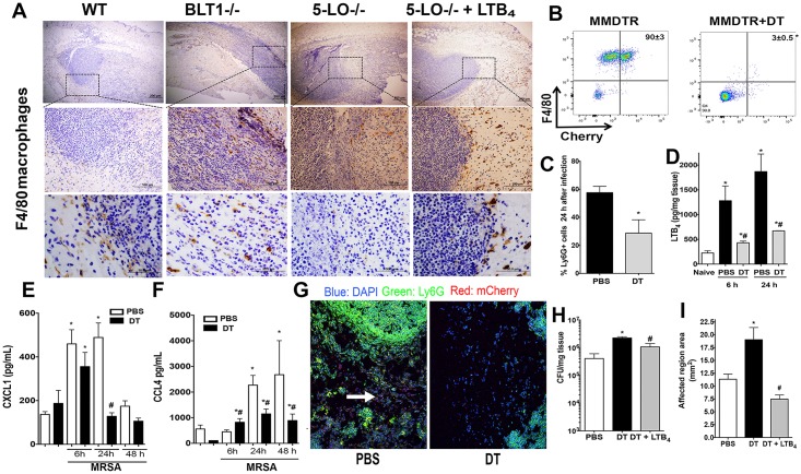
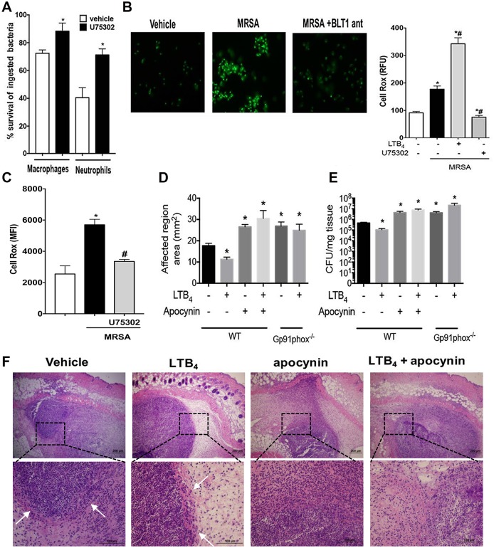
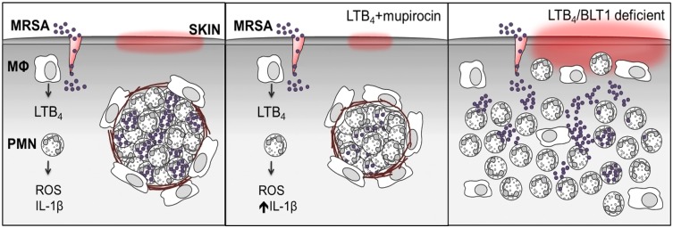
The early events that shape the innate immune response to restrain pathogens during skin infections remain elusive. Methicillin-resistant Staphylococcus aureus (MRSA) infection engages phagocyte chemotaxis, abscess formation, and microbial clearance. Upon infection, neutrophils and monocytes find a gradient of chemoattractants that influence both phagocyte direction and microbial clearance. The bioactive lipid leukotriene B4 (LTB4) is quickly (seconds to minutes) produced by 5-lipoxygenase (5-LO) and signals through the G protein-coupled receptors LTB4R1 (BLT1) or BLT2 in phagocytes and structural cells. Although it is known that LTB4 enhances antimicrobial effector functions in vitro, whether prompt LTB4 production is required for bacterial clearance and development of an inflammatory milieu necessary for abscess formation to restrain pathogen dissemination is unknown. We found that LTB4 is produced in areas near the abscess and BLT1 deficient mice are unable to form an abscess, elicit neutrophil chemotaxis, generation of neutrophil and monocyte chemokines, as well as reactive oxygen species-dependent bacterial clearance. We also found that an ointment containing LTB4 synergizes with antibiotics to eliminate MRSA potently. Here, we uncovered a heretofore unknown role of macrophage-derived LTB4 in orchestrating the chemoattractant gradient required for abscess formation, while amplifying antimicrobial effector functions.


Conflict of interest statement

The authors have declared that no competing interests exist.

Figures

Fig 1
Fig 1. MRSA infection induces LTB4 production in areas close to the abscess.
A) Detection of Alox5 and Ltb4r1 from the uninfected naïve skin and day 1 post-MRSA skin infection as assessed by qPCR. B) LTB4 EIA from skin biopsy homogenates from uninfected naïve samples, days 1 and 11 post-infection. C) Top panel: MALDI imaging for AA (red) determined by imaging mass spectrometry as described in the Methods. Bottom panel: H&E stained sections of naïve skin and day 1 post-MRSA skin infection. White dotted line indicates the edge of the skin tissue section. D) Expression of 5-LO at 40 X (left panel) and 100 X (middle panel) and 400 X (right) magnification in mice at day 1 after MRSA infection. The 5-LO is shown in brown and counterstained in blue. Data are representative of 2–3 mice. Dotted lines indicate abscess edges. E) Detection of 5-LO (red) and Ly6G (green) in the skin of MRSA-infected mice at 40 X (left panel), 100 X (middle panel) and 400 X (right panel). The inset represents 1000 X amplification. Colocalization between nucleus (DAPI) plus 5-LO is shown as purple, and 5-LO plus Ly6G is shown in yellow. Data are the mean ± SEM from 3–6 mice from 2–3 experiments. *p < 0.05 vs. naïve.
Fig 2
Fig 2. LTB4/BLT1 axis is essential to control MRSA skin infections.
A) The infection area was measured every other day for 9 days in wild-type (WT) C57BL/6 and BLT1-/- mice infected subcutaneously (s.c.) with MRSA and then treated daily or not treated with BLT1 antagonist U75302 (U7) or vehicle control. B) Bacterial loads (CFU counts) determined at days 1 and 9 after MRSA skin infection in C57BL/6, 5-LO-/-, BLT1-/-, and BLT1 antagonist-treated mice. C) Infection areas were determined as in (A) in the C57BL/6, and 5-LO-/- mice treated or not treated daily with LTB4 ointment. D) Bacterial load in the infected skin determined as in (B) in WT and 5-LO-/- mice treated or not treated with LTB4 ointment. E) Gram staining of Skin biopsies collected at day 1 post-MRSA skin infection from WT, BLT1-/-, 5-LO-/-, and 5-LO-/- mice treated topically with 3.37 × 10⁻⁶% LTB4 ointment. Gram staining to label gram-positive bacteria is shown in purple/brown. Top panels are 40 X, middle panels are 100 X, and bottom panels are 400 X magnification Insets represent 1000 X magnification. Black arrows indicate MRSA clusters. Images are representative of 3–5 mice. F) H&E stains from mice treated as in (E) and shown at 40 X (upper) and 100 X magnification. G) Thickness of Masson’s trichrome blue positive layer surrounding the abscess was measured with ImageJ software. In all cases, data represent the mean ± SEM from 3–4 mice. *p < 0.05 vs. WT. #p < 0.05 vs. 5-LO-/- treated with vehicle-control ointment.
Fig 3
Fig 3. LTB4 acts synergistically with mupirocin to increase MRSA clearance.
(A) Infection area measured every other day for 9 days post infection in WT mice infected with MRSA, s.c., and treated topically daily with vehicle-control ointment, 0.1% mupirocin ointment, LTB4 ointment, or mupirocin + LTB4 combination ointment. B) Bacterial load determined by CFU measured in the skin biopsy homogenates from mice treated as in (A) and collected at day 9 post-infection. C) Top: Representative bioluminescent MRSA detected in mice treated as in (A) using the in vivo animal imaging (IVIS Spectrum) detection system for the skin. Bottom: Bacterial load of bioluminescent MRSA in the skin of mice treated as in (A) using representative planar bioluminescent imaging (n = 5–6 mice/group). D) H&E stains from mice treated as in (A) and biopsies collected at day 1 post-MRSA skin infection. 40 X magnification and the white dotted lines indicate an abscess edge. E) (left) Masson’s trichrome blue staining from mice infected and treated with vehicle, LTB4, and LTB4 plus mupirocin for 24 h. The top is 40 X, the middle is 100 X and bottom is 400 X magnification. (right) The thickness of Masson’s trichrome blue positive layer surrounding the abscess was measured with ImageJ software as described in the Material and Methods. Data are the mean ± SEM of 4–5 mice from 1–2 experiments. *p < 0.05 vs. vehicle. **** p < 0.001 vs. vehicle.
Fig 4
Fig 4. LTB4 is required for neutrophil association in the abscess.
(A) Heat map of proteins involved in inflammatory immune response and resolution in WT and BLT1-/- mice infected with MRSA at day 1 and day 9 after infection measured using bead array multiplex. Proteins are listed on right-hand y-axis; grouped alphabetically in clades. Red indicates higher abundance whereas blue represents lower abundance. Each column for each condition represents a technical replicate (n = 4-5/group). B) Skin biopsy sections from WT, BLT1-/-, 5-LO-/-, and 5-LO-/- mice + LTB4 ointment were stained with anti-Ly6G/C antibody as described in the Material and Methods. Top panels are 40 X, and bottom panels are 400 X magnification. Neutrophil staining is shown in brown and counterstained in blue. C) Detection of Ly6G positive cells in the skin biopsies of mice infected and treated with the vehicle control or the ointment containing 0.001% BLT1 antagonist for 24 h using FACS analysis. D) Still frames of the intravital imaging from the EGFP-LysM mice infected with MRSA and treated with ointments containing vehicle control (untreated), 3.37 ng LTB4 ointment, or 0.001% BLT1 antagonist (U75302). After 24 hours, the mice were imaged by two-photon intravital microscopy for 30 minutes. E) Determination of the swarm size in WT EGFP-LysM mice treated or not with the LTB4 ointment using image J as described in the Material and Methods. F) Quantifications and track paths of mice represented in (C) that show the median velocity of the GFP+ cells. Track displacements of GFP+ cells and ratios of displacement/track durations were measured using FIJI (ImageJ) Trackpath plugin software and analyses were as described in the Material and Methods. Data are the mean ± SEM of 1000+ GFP+ cells representative of 3–4 mice. ****p < 0.0001 vs. untreated. White arrows indicate swarming.
Fig 5
Fig 5. Skin macrophages are necessary for LTB4 production and host defenses during MRSA skin infection.
A) Skin biopsy sections from WT, BLT1-/-, 5-LO-/-, and 5-LO-/- plus the LTB4 ointment treated mice stained with the anti-F4/80 antibody as described in Material and Methods. The top panels are 40 X, the middle panels are 400 X, and the bottom panels are 1000 X magnification. Macrophages stained brown, and the counterstain is blue. Black arrows indicate macrophages and the black dashed box is the amplified region. Images are representative of 3–5 mice. B) Depletion of skin macrophages in MMDTR mice were PBS-treated or DT-treated and infected with MRSA by subcutaneous injection. C) Percentages of Ly6G+ cells in MMDTR mice treated or not treated with DT, followed by MRSA infection for 24 hours, and examined by FACS analyses. D) LTB4 production in the skin of MMDTR treated or not treated with DT mice, followed by infection at the indicated time points as detected by EIA. E and F) Production of CXCL1 (E) and CCL4 (F) in the skin biopsies from MMDTR mice treated or not treated with DT, followed by MRSA skin infection for the indicated time points as detected by ELISA. G) Slides were stained using DAPI (blue), mCherry for macrophages (red), and FITC-labeled anti-Ly6G antibody (green). Images shown are at 200 X magnification and are representative of 3–5 mice/group. White arrows indicate mCherry+ stained macrophages. H) Bacterial loads of MMDTR mice treated or not treated with DT, infected with MRSA, and treated with LTB4 ointment for 24 hours as measured by CFU counts/mg tissue. I) Lesion sizes of MMDTR mice treated as in (F) and measured 24 hours after infection. Data are the mean ± SEM of 3–8 mice. *p < 0.05 vs. naïve. #p < 0.05 vs PBS-treated mice.
Fig 6
Fig 6. LTB4 promotes NADPH oxidase-mediated killing of MRSA.
A) Determination of bacterial killing in peritoneal macrophages and bone marrow-derived neutrophils from WT and BLT1-/- mice as described in the Material and Methods. B) (left) Determination of reactive oxygen species (ROS) production in macrophages from WT mice treated or not treated with the BLT1 antagonist, followed by challenge with MRSA for 60 minutes. (Right) Relative fluorescence units (RFU) levels of CellRox positive macrophages treated with vehicle control, LTB4, and BLT1 antagonist, followed by MRSA infection for 60 minutes. The RFU were determined as described in the Material and Methods. C) Mean fluorescence intensity (MFI) levels of CellRox positive bone marrow-derived neutrophils treated with vehicle control and BLT1 antagonist, followed by MRSA infection for 60 minutes. The MFI was determined as described in the Material and Methods. D) WT or gp91phox-/- mice were infected with MRSA via s.c. and treated topically daily with vehicle-control ointment, apocynin, LTB4 ointment, or apocynin + LTB4 combination ointment. Infection areas were measured 24 hours after infection, and the affected areas were measured as described in the Material and Methods. E) Bacterial loads of WT and Gp91phox-/- mice treated as in (D) and CFU were determined 24 hours after infection as described in the Material and Methods. F) H&E staining of mice treated and infected as in (D). The top panels are 4 X, and the bottom panels are 40 X magnification. Data are the mean ± SEM of 8–15 mice. *p < 0.05 vs. WT. White arrows indicate abscess edges. All abbreviations are defined in Figs 1 and 2 legends.
Fig 7
Fig 7. Summary of LTB4-promoted MRSA skin infection clearance.
Left panel: MRSA skin infection requires tissue-resident macrophages for optimal LTB4 production and neutrophil recruitment. LTB4 provides direction to neutrophils to contain bacteria and form an abscess surrounded by fibrous collagen. Middle panel: Combination ointment with LTB4 + the topical antibiotic, mupirocin, is more efficient at eliminating the infection. Right panel: Mice lacking LTB4/BLT1 fail to contain bacteria or develop an abscess. All abbreviations are defined in Figs 1 and 2 legends.

Comment in

  • Fatal chemoattraction.
    Bucci M. Bucci M. Nat Chem Biol. 2018 Oct;14(10):903. doi: 10.1038/s41589-018-0140-2. Nat Chem Biol. 2018. PMID: 30224689 No abstract available.

References

    1. Trividic M, Gauthier ML, Sparsa A, Ploy MC, Mounier M, Boulinguez S, et al. [Methi-resistant Staphylococcus aureus in dermatological practice: origin, risk factors and outcome]. Ann Dermatol Venereol. 2002;129(1 Pt 1):27–9. Epub 2002/04/09. . - PubMed
    1. Vayalumkal JV, Whittingham H, Vanderkooi O, Stewart TE, Low DE, Mulvey M, et al. Necrotizing pneumonia and septic shock: suspecting CA-MRSA in patients presenting to Canadian emergency departments. CJEM. 2007;9(4):300–3. . - PubMed
    1. Wong CH, Tan SH, Kurup A, Tan AB. Recurrent necrotizing fasciitis caused by methicillin-resistant Staphylococcus aureus. Eur J Clin Microbiol Infect Dis. 2004;23(12):909–11. Epub 2004/12/16. 10.1007/s10096-004-1237-y . - DOI - PubMed
    1. Miller LS, Cho JS. Immunity against Staphylococcus aureus cutaneous infections. Nat Rev Immunol. 2011;11(8):505–18. 10.1038/nri3010 . - DOI - PMC - PubMed
    1. Gessler P, Pretre R, Hohl V, Rousson V, Fischer J, Dahinden C. CXC-chemokine stimulation of neutrophils correlates with plasma levels of myeloperoxidase and lactoferrin and contributes to clinical outcome after pediatric cardiac surgery. Shock. 2004;22(6):513–20. . - PubMed

Publication types

MeSH terms