

Isolation and characterization of a highly pathogenic strain of Porcine enteric alphacoronavirus causing watery diarrhoea and high mortality in newborn piglets
- PMID: 30103259
- PMCID: PMC7168553
- DOI: 10.1111/tbed.12992
Isolation and characterization of a highly pathogenic strain of Porcine enteric alphacoronavirus causing watery diarrhoea and high mortality in newborn piglets
Abstract
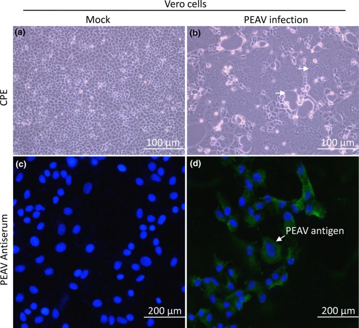
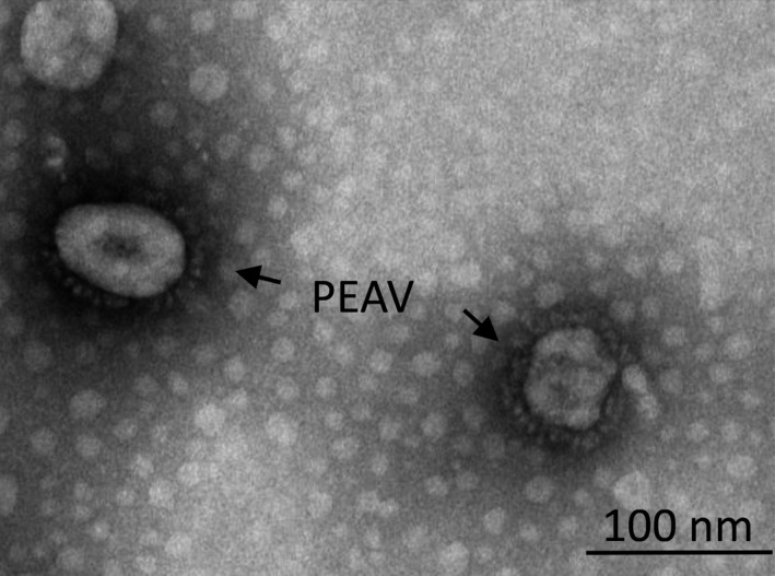
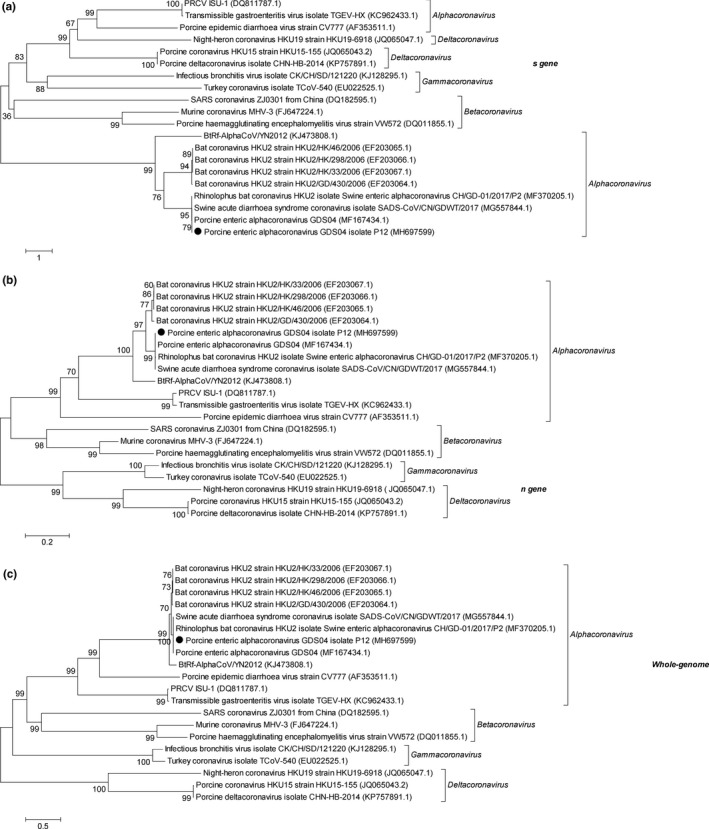
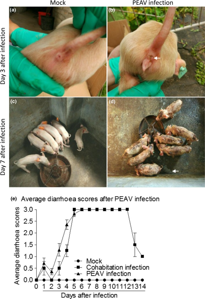
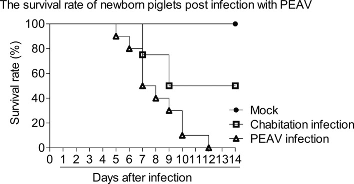
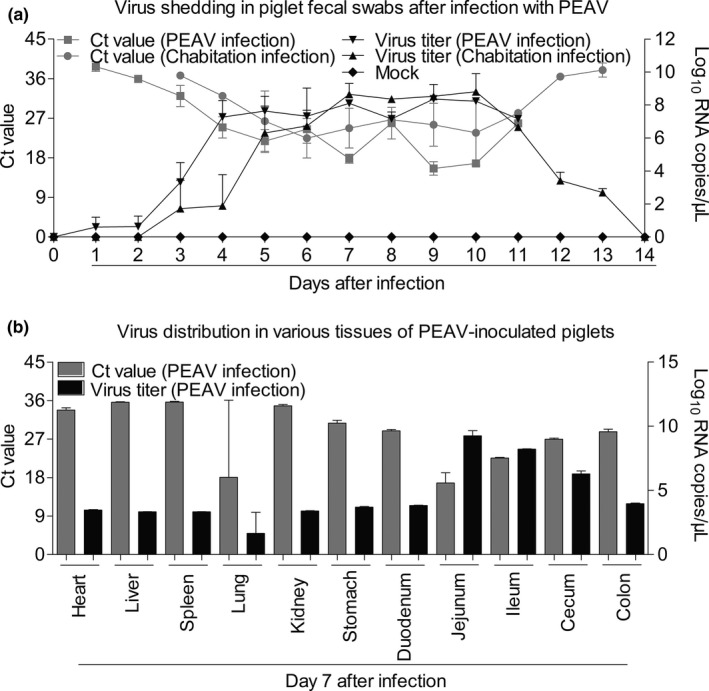
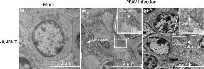
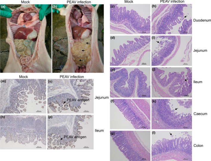
Porcine enteric alphacoronavirus (PEAV) was first discovered in China in February 2017, and the origin and virulence of this novel porcine coronavirus were not fully characterized. Here, we isolated a strain of PEAV, named GDS04 that is identified by immunofluorescence and typical crown-shaped particles observed with electron microscopy. Genomic analysis reveals that PEAV GDS04 shares a close relationship with SADS-CoV and SeACoV. Furthermore, newborn piglets orally challenged with PEAV GDS04 developed typical clinical symptoms as watery diarrhoea in neonatal piglets. Viral RNA was detected in faeces and various tissues of the infected piglets. Moreover, macroscopic and microscopic lesions in whole intestinal tract were observed, and viral antigen could be detected in the small intestines by immunohistochemical staining and electron microscopy. Importantly, the mortality rate of inoculated-newborn piglets was 100% and half of the cohabiting piglets died. Collectively, we demonstrate that PEAV is highly pathogenic in newborn piglets.
Keywords: Porcine enteric alphacoronavirus (PEAV); newborn piglets; pathogenicity.
© 2018 Blackwell Verlag GmbH.
Conflict of interest statement
The authors declare that they have no conflict of interest.
Figures
Similar articles
-
Attenuation and characterization of porcine enteric alphacoronavirus strain GDS04 via serial cell passage.Vet Microbiol. 2019 Dec;239:108489. doi: 10.1016/j.vetmic.2019.108489. Epub 2019 Nov 4. Vet Microbiol. 2019. PMID: 31767069 Free PMC article.
-
Discovery of a novel swine enteric alphacoronavirus (SeACoV) in southern China.Vet Microbiol. 2017 Nov;211:15-21. doi: 10.1016/j.vetmic.2017.09.020. Epub 2017 Sep 28. Vet Microbiol. 2017. PMID: 29102111 Free PMC article.
-
A New Bat-HKU2-like Coronavirus in Swine, China, 2017.Emerg Infect Dis. 2017 Sep;23(9):1607-9. doi: 10.3201/eid2309.170915. Epub 2017 Sep 17. Emerg Infect Dis. 2017. PMID: 28654418 Free PMC article.
-
Swine enteric alphacoronavirus (swine acute diarrhea syndrome coronavirus): An update three years after its discovery.Virus Res. 2020 Aug;285:198024. doi: 10.1016/j.virusres.2020.198024. Epub 2020 May 16. Virus Res. 2020. PMID: 32482591 Free PMC article. Review.
-
Emerging and re-emerging coronaviruses in pigs.Curr Opin Virol. 2019 Feb;34:39-49. doi: 10.1016/j.coviro.2018.12.001. Epub 2019 Jan 14. Curr Opin Virol. 2019. PMID: 30654269 Free PMC article. Review.
Cited by
-
Detection and Genetic Diversity of Porcine Coronavirus Involved in Diarrhea Outbreaks in Spain.Front Vet Sci. 2021 Feb 25;8:651999. doi: 10.3389/fvets.2021.651999. eCollection 2021. Front Vet Sci. 2021. PMID: 33718476 Free PMC article.
-
Emodin from Aloe inhibits Swine acute diarrhea syndrome coronavirus in cell culture.Front Vet Sci. 2022 Aug 18;9:978453. doi: 10.3389/fvets.2022.978453. eCollection 2022. Front Vet Sci. 2022. PMID: 36061121 Free PMC article.
-
Identification of ZDHHC17 as a Potential Drug Target for Swine Acute Diarrhea Syndrome Coronavirus Infection.mBio. 2021 Oct 26;12(5):e0234221. doi: 10.1128/mBio.02342-21. Epub 2021 Oct 26. mBio. 2021. PMID: 34700373 Free PMC article.
-
Interplay of swine acute diarrhoea syndrome coronavirus and the host intrinsic and innate immunity.Vet Res. 2025 Jan 9;56(1):5. doi: 10.1186/s13567-024-01436-1. Vet Res. 2025. PMID: 39789633 Free PMC article. Review.
-
Transcriptional Landscape of Vero E6 Cells during Early Swine Acute Diarrhea Syndrome Coronavirus Infection.Viruses. 2021 Apr 14;13(4):674. doi: 10.3390/v13040674. Viruses. 2021. PMID: 33919952 Free PMC article.
References
-
- Alsaad, K. O. , Hajeer, A. H. , Al Balwi, M. , Al Moaiqel, M. , Al Oudah, N. , Al Ajlan, A. , … Arabi, Y. M. (2018). Histopathology of Middle East respiratory syndrome coronovirus (MERS‐CoV) infection ‐ clinicopathological and ultrastructural study. Histopathology, 72, 516–524. 10.1111/his.13379 - DOI - PMC - PubMed
MeSH terms
Substances
Associated data
- Actions
- Actions
- Actions
Grants and funding
LinkOut - more resources
Full Text Sources
Other Literature Sources
Medical
