Review
. 2018 Aug 11;6(3):84.
doi: 10.3390/microorganisms6030084.

Diversity of Myxobacteria-We Only See the Tip of the Iceberg

Affiliations
Review

Diversity of Myxobacteria-We Only See the Tip of the Iceberg

Kathrin I Mohr. Microorganisms. .

Abstract

The discovery of new antibiotics is mandatory with regard to the increasing number of resistant pathogens. One approach is the search for new antibiotic producers in nature. Among actinomycetes, Bacillus species, and fungi, myxobacteria have been a rich source for bioactive secondary metabolites for decades. To date, about 600 substances could be described, many of them with antibacterial, antifungal, or cytostatic activity. But, recent cultivation-independent studies on marine, terrestrial, or uncommon habitats unequivocally demonstrate that the number of uncultured myxobacteria is much higher than would be expected from the number of cultivated strains. Although several highly promising myxobacterial taxa have been identified recently, this so-called Great Plate Count Anomaly must be overcome to get broader access to new secondary metabolite producers. In the last years it turned out that especially new species, genera, and families of myxobacteria are promising sources for new bioactive metabolites. Therefore, the cultivation of the hitherto uncultivable ones is our biggest challenge.

Keywords: diversity; myxobacteria; new antibiotics; secondary metabolites; uncultured.


Conflict of interest statement

The authors declare no conflict of interest.

Figures

Figure 1
Figure 1
Monophyletic order Myxococcales (delta-proteobacteria), suborders, families, and genera of myxobacteria (status May 2018). The number of species within the genera is mentioned in brackets (original graphic from Corinna Wolf, modified by K. I. Mohr).
Figure 2
Figure 2
Variation of myxobacterial fruiting bodies. Genus/species, strain designation, (agar medium) are mentioned. (a) Myxococcus xanthus Mxx42 (P); (b) Cystobacter ferrugineus Cbfe48 (VY/2); (c) Archangium sp. Ar7747 (VY/2); (d) Chondromyces sp. (Stan 21 with filter); (e) Sorangium sp. Soce 1462 degrading filter paper on Stan 21 agar; (f) Polyangium sp. Pl3323 (VY/2); (g) Cystobacter fuscus Cbf18 (VY/2); (h) Corallococcus coralloides Ccc379 (VY/2).
Figure 3
Figure 3
Sorangium sp. strain Soce 1014, an ambruticin-producer, swarming on VY/2-agar and the structure of ambruticin A, the first secondary metabolite which was isolated and described from myxobacteria.
Figure 4
Figure 4
Neighbour joining tree with myxobacterial type strains shows the phylogenetic position of strain WY75, cultivated from ginger foundation soil, within the Sorangiineae suborder. Comparison of 16S rRNA sequences revealed only 87.4% similarity to the next myxobacterial type strain S. amylolyticus. Accession numbers are in brackets. Bar, 0.1 substitutions per nucleotide position.
Figure 5
Figure 5
Common isolation procedure for myxobacteria: Soil/environmental sample is placed on a. Stan 21 with filter paper to enrich cellulose decomposing strains and b. on water agar with E. coli bait (cross) for predators. Numerous transfers of fruiting bodies/swarm edge material to fresh plates are necessary to purify myxobacteria.
Figure 6
Figure 6
Myxobacterial cultures isolated from Kiritimati sand (a,d) and German compost (bf) modified from Mohr et al. [9]. (a) Corallococcus (Myxococcus) macrosporus, (b) Corallococcus sp., (c) Myxococcus sp. (d) Archangium gephyra, (e) Corallococcus sp., (f) Polyangium fumosum.
Figure 7
Figure 7
(a) Brockenfeld high moor; (b) fen Am Sandbeek; (c) Brockenfeld high moor scarp. Isolated Corallococcus sp. strains from moors (d) strain B19, (e) strain B2t-1. (f) Sorangium cellulosum (orange) on a raw culture plate (Stan 21 with filter) inoculated with soil material from moor. Pictures are from Mohr et al. (2017) and modified [2].
Figure 8
Figure 8
Part of a distance tree showing some myxobacterial type strains, some representative clone sequences and one representative culture sequence from our study [2] as well as sequences from uncultivated myxobacteria from other studies. Red: clones from Brockenfeld high moor; blue: clones from fen Am Sandbeek; green: representative culture from Brockenfeld high moor. Accession numbers are given in brackets. Origin of sequences from uncultured myxobacteria are mentioned. Bar, 0.1 substitutions per nucleotide position.
Figure 9
Figure 9
Marine myxobacterial type strains on agar plates. (a) Haliangium tepidum (DSM 14436T) on VY/2SWS, (b) Enhygromyxa salina (DSM 15217T) on VY/4SWS, (c) Pseudenhygromyxa salsuginis (DSM 21377T) on 1102, (d) Plesiocystis pacifica (DSM 14875T) on VY/2SWS.
Figure 10
Figure 10
Neighbour joining tree of myxobacterial type strains (16S rRNA-genes), some representative clones from the MMC-cluster (blue) and the next cultivated relative Sandaracinus amylolyticus. Genera isolated from marine environment are in green blue. Suborders of the order Myxococcales, origin of clones, and accession numbers are shown. Bar, 0.1 substitutions per nucleotide position.
Figure 11
Figure 11
Neighbour joining tree of some myxobacterial type strains (16S rRNA-genes) and some representative clones. All clones show highest relationship to the next cultivated relative A. dehalogenans. All families of the Cystobacterineae suborder, origin of samples, and accession numbers of clones/cultures are shown. Bar, 0.1 substitutions per nucleotide position.
Figure 12
Figure 12
Moderately thermophilic strains of Sorangium on VY/2 agar. (a) GT47 and (b) GT 41, isolated by Dr. K. Gerth.
Figure 13
Figure 13
Neighbour joining tree of some myxobacterial type strains (16S rRNA-genes) and cultures (in bold) from hot springs (AB246767-AB246770, AB246772) [117] and alkaline hot spring, all from Japan. Suborders of the order Myxococcales, origin of samples, and accession numbers are shown. Bar, 0.1 substitutions per nucleotide position.

Similar articles

Cited by

References

    1. Dawid W. Biology and global distribution of myxobacteria in soils. FEMS Microbiol. Rev. 2000;24:403–427. doi: 10.1111/j.1574-6976.2000.tb00548.x. - DOI - PubMed
    1. Mohr K.I., Zindler T., Wink J., Wilharm E., Stadler M. Myxobacteria in high moor and fen: An astonishing diversity in a neglected extreme habitat. MicrobiologyOpen. 2017;6 doi: 10.1002/mbo3.464. - DOI - PMC - PubMed
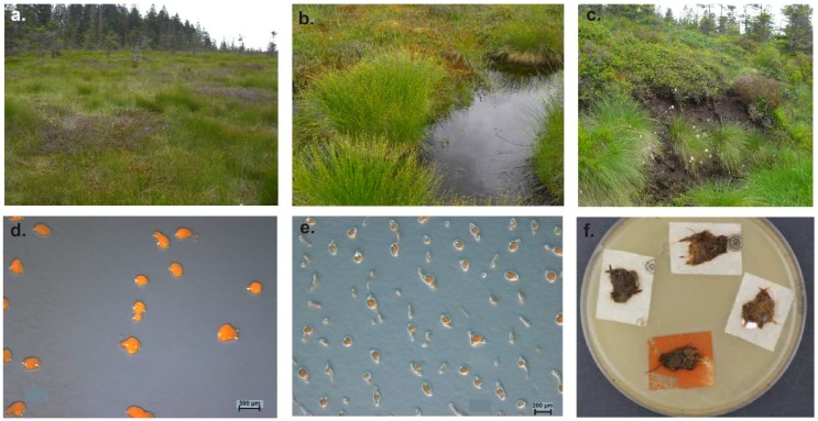
    1. Dawid W. Myxobakterien in ungestörten Hochmooren des Hohen Venn (Hautes Fagnes, Belgien) Syst. Appl. Microbiol. 1984;5:555–563. doi: 10.1016/S0723-2020(84)80013-2. - DOI
    1. Fudou R., Jojima Y., Iizuka T., Yamanaka S. Haliangium ochraceum gen. nov., sp. nov. and Haliangium tepidum sp. nov.: Novel moderately halophilic myxobacteria isolated from coastal saline environments. J. Gen. Appl. Microbiol. 2002;48:109–116. doi: 10.2323/jgam.48.109. - DOI - PubMed
    1. Iizuka T., Jojima Y., Fudou R., Hiraishi A., Ahn J.W., Yamanaka S. Plesiocystis pacifica gen. nov., sp. nov., a marine myxobacterium that contains dihydro-genated menaquinone, isolated from the Pacific coasts of Japan. IJSEM. 2003;53:189–195. doi: 10.1099/ijs.0.02418-0. - DOI - PubMed

LinkOut - more resources