. 2018 Aug 12;5(3):66.
doi: 10.3390/bioengineering5030066.

A Precisely Flow-Controlled Microfluidic System for Enhanced Pre-Osteoblastic Cell Response for Bone Tissue Engineering

Affiliations

A Precisely Flow-Controlled Microfluidic System for Enhanced Pre-Osteoblastic Cell Response for Bone Tissue Engineering

Eleftheria Babaliari et al. Bioengineering (Basel). .

Abstract

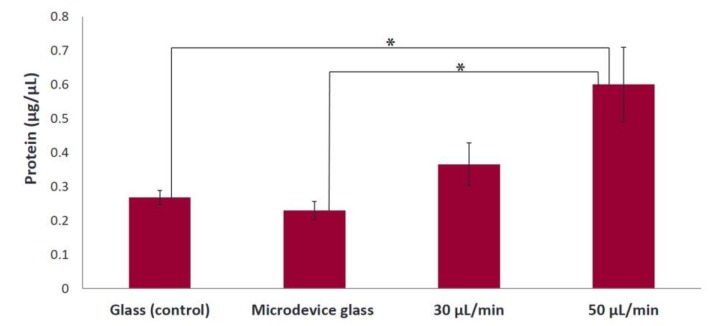
Bone tissue engineering provides advanced solutions to overcome the limitations of currently used therapies for bone reconstruction. Dynamic culturing of cell-biomaterial constructs positively affects the cell proliferation and differentiation. In this study, we present a precisely flow-controlled microfluidic system employed for the investigation of bone-forming cell responses cultured on fibrous collagen matrices by applying two flow rates, 30 and 50 μL/min. We characterized the collagen substrates morphologically by means of scanning electron microscopy, investigated their viscoelastic properties, and evaluated the orientation, proliferation and osteogenic differentiation capacity of pre-osteoblastic cells cultured on them. The cells are oriented along the direction of the flow at both rates, in contrast to a random orientation observed under static culture conditions. The proliferation of cells after 7 days in culture was increased at both flow rates, with the flow rate of 50 μL/min indicating a significant increase compared to the static culture. The alkaline phosphatase activity after 7 days increased at both flow rates, with the rate of 30 μL/min indicating a significant enhancement compared to static conditions. Our results demonstrate that precisely flow-controlled microfluidic cell culture provides tunable control of the cell microenvironment that directs cellular activities involved in bone regeneration.

Keywords: MC3T3-E1 pre-osteoblasts; cell orientation; collagen; microfluidics; osteogenic differentiation.


Conflict of interest statement

The authors declare no conflict of interest.

Figures

Figure 1
Figure 1
Illustration of the microfluidic system with precisely controlled flow. The microfluidic system is composed of a pressure pump (A) connected with a nutrient reservoir (B), a precisely controlled flow sensor (C), a chamber (D) including the microfluidic devices (E, F or G) and a waste reservoir (H).
Figure 2
Figure 2
Morphology of MC3T3-E1 cells inside the flow perfusion culture system using gelatin substrates under static conditions (A), and under flow conditions applying 30 (B,C), and 50 μL/min (D,E) visualized by optical microscopy (ten-fold magnification, the scale bars represent 50 μm). The black arrows represent the direction of the flow. Directionality histograms and tables with the statistics generated by means of the Fiji ImageJ plug-in “Directionality” [25] are presented directly under the optical microscopy images (AE).
Figure 3
Figure 3
Normalized levels of collagen secreted by MC3T3-E1 cells on gelatin substrates after 7 days of culture under static conditions (glass and microdevice glass) and under flow conditions (flow rates of 30 and 50 μL/min). A *p value of < 0.05 was considered significant.
Figure 4
Figure 4
SEM images showing the morphology of fibrous collagen networks (A); SEM image of a sample cross-section showing the fibrous collagen scaffold prepared on a glass substrate (B).
Figure 5
Figure 5
Linear viscoelastic response measured by a Dynamic frequency sweep test for a 3 mg/mL collagen hydrogel at 37 °C. The elastic modulus, G’, is represented by solid symbols and the viscous modulus, G’’, by open ones.
Figure 6
Figure 6
SEM images show the morphology of MC3T3-E1 cells after 1 day of culture spreading onto fibrous collagen; images with different magnifications, 1000× (A) and 3000× (B) indicate a fully flattened pre-osteoblastic cell morphology and cell protrusions, respectively.
Figure 7
Figure 7
Morphology of MC3T3-E1 cells inside the flow perfusion culture system using fibrous collagen substrates under static conditions (A), and under flow conditions applying 30 (B,C), and 50 μL/min (D,E) using optical microscopy (ten-fold magnification, the scale bar represents 50 μm). The black arrows represent the direction of the flow. Directionality histograms and tables with the statistics generated by means of the Fiji ImageJ plug-in “Directionality” [25] are presented directly under the optical microscopy images (AE).
Figure 8
Figure 8
Amount of cells expressed as total protein concentration after 7 days of culture on fibrous collagen substrates under static (glass and microdevice glass) and flow conditions (flow rates of 30 and 50 μL/min). A *p value of < 0.05 was considered significant.
Figure 9
Figure 9
Normalized ALP activity of MC3T3-E1 cells on fibrous collagen substrates after 7 days of culture under static (glass and microdevice glass) and flow conditions (flow rates of 30 and 50 μL/min) in the presence of osteogenic medium. A *p value of < 0.05 was considered significant.

References

    1. Danilevicius P., Rezende R.A., Pereira F.D., Selimis A., Kasyanov V., Noritomi P.Y., da Silva J.V., Chatzinikolaidou M., Farsari M., Mironov V. Burr-like, laser-made 3D microscaffolds for tissue spheroid encagement. Biointerphases. 2015;10:021011. doi: 10.1116/1.4922646. - DOI - PubMed
    1. Wang Y.L., Pelham R.J. Preparation of a flexible, porous polyacrylamide substrate for mechanical studies of cultured cells. Method Enzymol. 1998;298:489–496. - PubMed
    1. Kim B.S., Putnam A.J., Kulik T.J., Mooney D.J. Optimizing seeding and culture methods to engineer smooth muscle tissue on biodegradable polymer matrices. Biotechnol. Bioeng. 1998;57:46–54. doi: 10.1002/(SICI)1097-0290(19980105)57:1<46::AID-BIT6>3.0.CO;2-V. - DOI - PubMed
    1. Vunjak-Novakovic G., Obradovic B., Martin I., Bursac P.M., Langer R., Freed L.E. Dynamic cell seeding of polymer scaffolds for cartilage tissue engineering. Biotechnol. Progr. 1998;14:193–202. doi: 10.1021/bp970120j. - DOI - PubMed
    1. Granet C., Laroche N., Vico L., Alexandre C., Lafage-Proust M.H. Rotating-wall vessels, promising bioreactors for osteoblastic cell culture: comparison with other 3D conditions. Med. Biol. Eng. Comput. 1998;36:513–519. doi: 10.1007/BF02523224. - DOI - PubMed

LinkOut - more resources