Mesenchymal stem cell transplantation improves chronic colitis-associated complications through inhibiting the activity of toll-like receptor-4 in mice
- PMID: 30103680
- PMCID: PMC6090774
- DOI: 10.1186/s12876-018-0850-7
Mesenchymal stem cell transplantation improves chronic colitis-associated complications through inhibiting the activity of toll-like receptor-4 in mice
Abstract
Background: A variety of extra-intestinal manifestations (EIMs), including hepatobiliary complications, are associated with inflammatory bowel disease (IBD). Mesenchymal stem cells (MSCs) have been shown to play a potential role in the therapy of IBD. This study was designed to investigate the effect and mechanism of MSCs on chronic colitis-associated hepatobiliary complications using mouse chronic colitis models induced by dextran sulfate sodium (DSS).
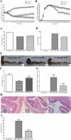
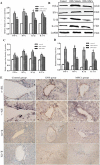
Methods: DSS-induced mouse chronic colitis models were established and treated with MSCs. Severity of colitis was evaluated by disease activity index (DAI), body weight (BW), colon length and histopathology. Serum lipopolysaccharide (LPS) levels were detected by limulus amebocyte lysate test (LAL-test). Histology and liver function of the mice were checked correspondingly. Serum LPS levels and bacterial translocation of mesenteric lymph nodes (MLN) were detected. Pro-inflammatory cytokines including tumor necrosis factor-α (TNF-α), interferon-γ (IFN-γ), interleukin-1β (IL-1β), interleukin-17A (IL-17A), Toll receptor 4 (TLR4), TNF receptor-associated factor 6 (TRAF6) and nuclear factor kappa B (NF-κB) were detected by immunohistochemical staining, western blot analysis and real-time PCR, respectively.
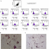
Results: The DSS-induced chronic colitis model was characterized by reduced BW, high DAI, worsened histologic inflammation, and high levels of LPS and E. coli. Liver histopathological lesions, impaired liver function, enhanced proteins and mRNA levels of TNF-α, IFN-γ, IL-1β, IL-17A, TLR4, TRAF6 and NF-κB were observed after DSS administration. MSCs transplantation markedly ameliorated the pathology of colon and liver by reduction of LPS levels and proteins and mRNA expressions of TNF-α, IFN-γ, IL-1β, IL-17A, TLR4, TRAF6 and NF-κB.
Conclusions: MSCs can improve chronic colitis-associated hepatobiliary complications, probably by inhibition of enterogenous endotoxemia and hepatic inflammation through LPS/TLR4 pathway. MSCs may represent a novel therapeutic approach for chronic colitis-associated hepatobiliary complications.
Keywords: Chronic colitis; Hepatobiliary complications; Lipopolysaccharide; Mesenchymal stem cells; Toll receptor 4.
Conflict of interest statement
Not applicable.
The authors declare that they had no competing interests.
Springer Nature remains neutral with regard to jurisdictional claims in published maps and institutional affiliations.
Figures


Similar articles
-
Human umbilical cord blood mesenchymal stem cells reduce colitis in mice by activating NOD2 signaling to COX2.Gastroenterology. 2013 Dec;145(6):1392-403.e1-8. doi: 10.1053/j.gastro.2013.08.033. Epub 2013 Aug 21. Gastroenterology. 2013. PMID: 23973922
-
Toll-like receptor 2 monoclonal antibody or/and Toll-like receptor 4 monoclonal antibody increase counts of Lactobacilli and Bifidobacteria in dextran sulfate sodium-induced colitis in mice.J Gastroenterol Hepatol. 2012 Jan;27(1):110-9. doi: 10.1111/j.1440-1746.2011.06839.x. J Gastroenterol Hepatol. 2012. PMID: 21722182
-
Zhikang Capsule ameliorates dextran sodium sulfate-induced colitis by inhibition of inflammation, apoptosis, oxidative stress and MyD88-dependent TLR4 signaling pathway.J Ethnopharmacol. 2016 Nov 4;192:236-247. doi: 10.1016/j.jep.2016.07.055. Epub 2016 Jul 21. J Ethnopharmacol. 2016. PMID: 27452656
-
Toll-like receptor 4 (TLR4) antagonists as potential therapeutics for intestinal inflammation.Indian J Gastroenterol. 2021 Feb;40(1):5-21. doi: 10.1007/s12664-020-01114-y. Epub 2021 Mar 5. Indian J Gastroenterol. 2021. PMID: 33666891 Free PMC article. Review.
-
CellSaic, A Cell Aggregate-Like Technology Using Recombinant Peptide Pieces for MSC Transplantation.Curr Stem Cell Res Ther. 2019;14(1):52-56. doi: 10.2174/1574888X13666180912125157. Curr Stem Cell Res Ther. 2019. PMID: 30207243 Free PMC article. Review.
Cited by
-
Stem cell therapy as a promising strategy in necrotizing enterocolitis.Mol Med. 2022 Sep 6;28(1):107. doi: 10.1186/s10020-022-00536-y. Mol Med. 2022. PMID: 36068527 Free PMC article. Review.
-
A novel therapeutic approach for inflammatory bowel disease by exosomes derived from human umbilical cord mesenchymal stem cells to repair intestinal barrier via TSG-6.Stem Cell Res Ther. 2021 May 29;12(1):315. doi: 10.1186/s13287-021-02404-8. Stem Cell Res Ther. 2021. PMID: 34051868 Free PMC article.
-
Impact of miR-223-3p and miR-2909 on inflammatory factors IL-6, IL-1ß, and TNF-α, and the TLR4/TLR2/NF-κB/STAT3 signaling pathway induced by lipopolysaccharide in human adipose stem cells.PLoS One. 2019 Feb 26;14(2):e0212063. doi: 10.1371/journal.pone.0212063. eCollection 2019. PLoS One. 2019. PMID: 30807577 Free PMC article.
-
Therapeutic potential of mesenchymal stem/stromal cells (MSCs)-based cell therapy for inflammatory bowel diseases (IBD) therapy.Eur J Med Res. 2023 Jan 27;28(1):47. doi: 10.1186/s40001-023-01008-7. Eur J Med Res. 2023. PMID: 36707899 Free PMC article. Review.
-
Protective effect and mechanism of mesenchymal stem cells on heat stroke induced intestinal injury.Exp Ther Med. 2020 Oct;20(4):3041-3050. doi: 10.3892/etm.2020.9051. Epub 2020 Jul 27. Exp Ther Med. 2020. PMID: 32855671 Free PMC article.
References
-
- Yang X, Meng J. Hepatopancreatobiliary complications among patients with inflammatory bowel disease. Chin Gen Pract. 2011;14:2398–2401.
MeSH terms
Substances
LinkOut - more resources
Full Text Sources
Other Literature Sources
Medical

