Randomized Controlled Trial
. 2018 Aug 13;15(1):229.
doi: 10.1186/s12974-018-1262-x.

RAS modulation prevents progressive cognitive impairment after experimental stroke: a randomized, blinded preclinical trial

Affiliations
Randomized Controlled Trial

RAS modulation prevents progressive cognitive impairment after experimental stroke: a randomized, blinded preclinical trial

Heba A Ahmed et al. J Neuroinflammation. .

Abstract

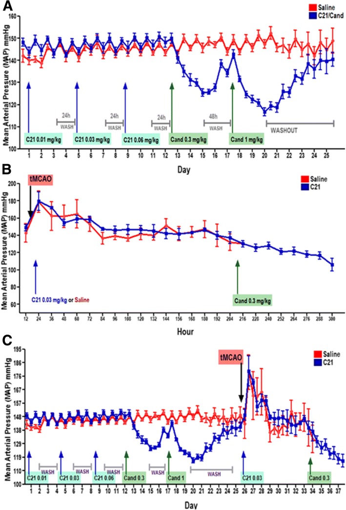
Background: With the aging population, the prevalence and incidence of cerebrovascular disease will continue to rise, as well as the number of individuals with vascular cognitive impairment/dementia (VCID). No specific FDA-approved treatments for VCID exist. Although clinical evidence supports that angiotensin receptor blockers (ARBs) prevent cognitive decline in older adults, whether ARBs have a similar effect on VCID after stroke is unknown. Moreover, these agents reduce BP, which is undesirable in the acute stroke period, so we believe that giving C21 in this acute phase or delaying ARB administration would enable us to achieve the neurovascular benefits without the risk of unintended and potentially dangerous, acute BP lowering.

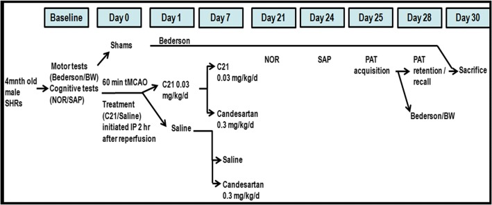
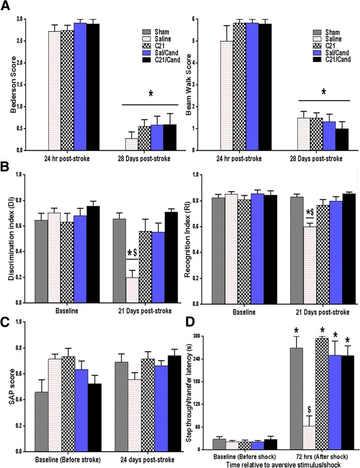
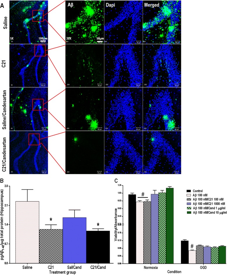
Methods: The aim of our study was to determine the impact of candesartan (ARB) or compound-21 (an angiotensin type 2 receptor--AT2R--agonist) on long-term cognitive function post-stroke, in spontaneously hypertensive rats (SHRs). We hypothesized that AT2R stimulation, either directly with C21, or indirectly by blocking the angiotensin type 1 receptor (AT1R) with candesartan, initiated after stroke, would reduce cognitive impairment. Animals were subjected to a 60-min transient middle cerebral artery occlusion and randomly assigned to either saline/C21 monotherapy, for the full study duration (30 days), or given sequential therapy starting with saline/C21 (7 days) followed by candesartan for the remainder of the study (21 days). Outcome measures included sensorimotor/cognitive-function, amyloid-β determination, and histopathologic analyses.

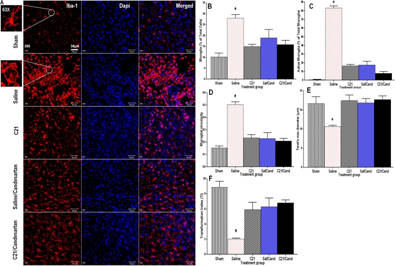
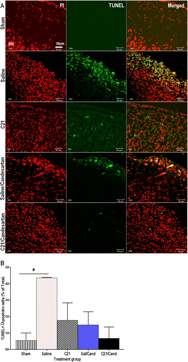
Results: Treatment with RAS modulators effectively preserved cognitive function, reduced cytotoxicity, and prevented chronic-reactive microgliosis in SHRs, post-stroke. These protective effects were apparent even when treatment was delayed up to 7 days post-stroke and were independent of blood pressure and β-amyloid accumulation.

Conclusion: Collectively, our findings demonstrate that RAS modulators effectively prevent cognitive impairment after stroke, even when treatment is delayed.

Keywords: Angiotensin modulators; Cognitive-impairment; Hypertension; Stroke.


Conflict of interest statement

All experimental procedures and animal protocols were approved by the Institutional Animal Care and Use Committee (IACUC) of Charlie Norwood VA Medical Center, Augusta, GA, USA.

Not applicable

Drugs were supplied from Astra-Zeneca and Vicore Pharma. The authors declared that they have no competing interests.

Springer Nature remains neutral with regard to jurisdictional claims in published maps and institutional affiliations.

Figures

Fig. 1
Fig. 1
Schematic time-line and description of the experimental design. This was a long-term 2 × 2, randomized, double-blind preclinical trial aimed to study the impact of RAS modulation, after stroke, on long-term cognitive function in spontaneously hypertensive rats (SHRs)
Fig. 2
Fig. 2
Compound 21 had no effect on blood pressure in SHRs after a 60-min tMCAO. a Mean arterial pressure (MAP), as measured by telemetry, showed no effect of C21 on BP, in unstroked animals. b There was an increase in MAP from baseline after transient middle cerebral artery occlusion (tMCAO) and this was not affected by C21 administration 2 h after reperfusion, and the average BP trend was similar for both C21 and saline-treated animals. c Figure depicting complete MAP trend, before and after tMCAO, with the effect of treatments administered at different points in time
Fig. 3
Fig. 3
RAS modulation improved learning and preserved spatial, non-spatial working, and long-term reference memories despite lack of effect on long-term motor recovery post-stroke. a All animals showed a significant recovery of sensorimotor function, as measured by Bederson (C21 × candesartan × time interaction F(1,48) = 0.22, P = 0.6384) and Beam walk (C21 × candesartan × time interaction F(1,31) = 0.41, P = 0.5278), at 28 days post-stroke, irrespective of their assigned treatment group. On the other hand, b for the novel object recognition (NOR) test, saline-treated animals showed a significant reduction in discrimination index (DI) and recognition index (RI), compared to their baseline values as well as to all other groups post-stroke, while sham and C21/candesartan-treated animals retained their ability to recognize the novel object during the preference trial indicating preserved non-spatial working memory (group × time interaction F(1,30) = 14.58, P < 0.0001). c Spontaneous alternation performance (SAP) was also noticeably better for these animals with plots of the means from baseline to 24 days showing distinctive changes within each group, shams, and candesartan/C21-treated groups having either no change from baseline or an increased SAP, while the saline group showed a clear decrease in SAP from 0 to 24 days indicating worsened spatial working memory (C21 × candesartan × time interaction F(1,17) = 0.12, P = 0.7314). d On the passive avoidance test (PAT), sham animals and those treated with C21/candesartan showed significantly enhanced step-through latency, indicating better memory retention and recall of the association between properties of the chamber and the foot shock, compared to their saline-treated counterparts. (group × time interaction F(1,39) = 19.00, P < 0.0001). For all tests n = 5–10 animals/group, error bars indicate SEM. Statistical significance is denoted by *P < 0.0001 for post hoc pair-wise comparisons from baseline and #P < 0.0001 for post hoc pair-wise comparisons “between groups” post-stoke, using the Bonferroni-adjusted alpha
Fig. 4
Fig. 4
RAS modulation prevented Aβ42-mediated cytotoxicity in HBECs and C21 prevented hippocampal Aβ42 accumulation in SHRs post-stroke. a Representative 5× and 20× immunofluorescent Aβ/green-stained images of SHR hippocampus with blue/dapi nuclear staining, scale bar represents 100 and 50 μm for 5× and 20× images, respectively. b An Aβ1–42 ELISA analysis showed that animals treated with C21, for the first 7 days, had markedly lower hippocampal concentrations of Aβ1–42 at 30 days post-stroke than those treated with saline. (C21 × candesartan interaction F(1,18) = 1.30, P = 0.2719, n = 5–10 animals/group. c Cell viability (MTT conversion) was significantly reduced in cultured HBECs incubated with Aβ1–42 compared with untreated controls, under similar conditions. This cytotoxicity was prevented when cells were co-treated with candesartan and higher dose C21 (condition by Aβ/C21/candesartan group interaction F(5,89) = 5.68, P = 0.0002, n = 6–12 wells/group, error bars indicate SEM). Statistical significance for post hoc comparisons between groups using Tukey’s multiple comparison procedure are denoted by *P < 0.05 to indicate a difference from saline and #P < 0.05 to indicate a difference from all other treatment groups
Fig. 5
Fig. 5
RAS modulation reduced microglial accumulation, offset sustained activation and prevented chronic reactive microgliosis, in SHRs post-stroke. a Representative immunofluorescent images depicting microglial morphology/ramification status changes as illustrated Iba-1 positive (red stained) microglia with blue dapi nuclear staining, scale bar represents 50 and 20 μm for 20× and 63× images, respectively. Hypertensive controls (saline) subjected to tMCAO had, in the ischemic hemisphere, a significantly higher proportion of b total microglia (F(4,14) = 13.18, P < 0.05) and c active, inflammatory microglia (F(4,14) = 249.1, P < 0.0001) at 30 days post-stroke relative to shams and C21/candesartan-treated animals. These saline-treated controls also showed d increased circularity (F(4,14) = 33.52, P < 0.0001), e reduced Feret’s maximum diameter (F(4,14) = 7.314, P < 0.05), and f reduced transformation index (TI) (F(4,14) = 14.02, P < 0.0001), all consistent with a transformation of microglia from ramified to reactive amoeboid forms, at 30 days post-stroke relative to both sham and C21/candesartan-treated animals. However, C21 and candesartan-treated animals showed indices not much different from those of shams (n = 3–4 animals/group, error bars indicate SEM). Statistical significance for post hoc comparisons between groups using Tukey’s multiple comparison procedure are denoted by *P < 0.05 for differences in proportion of total microglia and Feret’s maximum diameter and *P < 0.0001 for differences in all other parameters. Feret’s (maximum) diameter, a measure of cell length, is the greatest distance between any two points along the cell perimeter [51]. Circularity = 4π × [cell area (μm2)]/[cell perimeter (μm)]2 is unitless value that ranges from 0 (infinitely elongated)-1 (perfect circle) [51]. Transformation index (TI) = [perimeter of cell (μm)]2/4π [cell area(μm2)] [51]
Fig. 6
Fig. 6
RAS modulation reduced apoptotic cell death post-stroke. a Representative 20× immunofluorescent images depicting TUNEL positive (green stained) cortical cells with red propidium iodide nuclear staining, scale bar 50 μm. b Animals subjected to tMCAO had a significantly greater number of tunnel positive cells in the ischemic hemisphere than shams F(4,7) = 4.373, P = 0.0437. Cell death was greatly reduced in animals treated long-term with C21 and candesartan (n = 3–4 animals/group, error bars indicate SEM). Statistical significance for post hoc comparisons between groups using Tukey’s multiple comparison procedure are denoted by *P < 0.05 to indicate a difference from unstroked/sham animals

References

    1. Yang Y, Kimura-ohba S, Thompson J, Gary A. Rodent models of vascular cognitive impairment. Transl Stroke Res. 2016;7:407–414. doi: 10.1007/s12975-016-0486-2. - DOI - PMC - PubMed
    1. Iadecola C. The pathology of vascular dementia. Neuron. 2013;80:844–866. doi: 10.1016/j.neuron.2013.10.008. - DOI - PMC - PubMed
    1. Delavaran H, Jönsson A-C, Lövkvist H, Iwarsson S, Elmståhl S, Norrving B, et al. Cognitive function in stroke survivors: a 10-year follow-up study. Acta Neurol Scand. 2017;136:187–194. doi: 10.1111/ane.12709. - DOI - PubMed
    1. Gorelick P, Scuteri A, Black S. Vascular contributions to cognitive impairment and dementia: a statement for healthcare professionals from the American Heart Association/American Stroke Association. Stroke. 2011;42:2672–2713. doi: 10.1161/STR.0b013e3182299496. - DOI - PMC - PubMed
    1. Rouch L, Cestac P, Hanon O, Cool C, Helmer C, Bouhanick B, et al. Antihypertensive drugs, prevention of cognitive decline and dementia: a systematic review of observational studies, randomized controlled trials and meta-analyses, with discussion of potential mechanisms. CNS Drugs. 2015;29:113–130. doi: 10.1007/s40263-015-0230-6. - DOI - PubMed

Publication types

MeSH terms