. 2018 Aug 13;6(1):141.
doi: 10.1186/s40168-018-0524-2.

The tick endosymbiont Candidatus Midichloria mitochondrii and selenoproteins are essential for the growth of Rickettsia parkeri in the Gulf Coast tick vector

Affiliations

The tick endosymbiont Candidatus Midichloria mitochondrii and selenoproteins are essential for the growth of Rickettsia parkeri in the Gulf Coast tick vector

Khemraj Budachetri et al. Microbiome. .

Abstract

Background: Pathogen colonization inside tick tissues is a significant aspect of the overall competence of a vector. Amblyomma maculatum is a competent vector of the spotted fever group rickettsiae, Rickettsia parkeri. When R. parkeri colonizes its tick host, it has the opportunity to dynamically interact with not just its host but with the endosymbionts living within it, and this enables it to modulate the tick's defenses by regulating tick gene expression. The microbiome in A. maculatum is dominated by two endosymbiont microbes: a Francisella-like endosymbiont (FLE) and Candidatus Midichloria mitochondrii (CMM). A range of selenium-containing proteins (selenoproteins) in A. maculatum ticks protects them from oxidative stress during blood feeding and pathogen infections. Here, we investigated rickettsial multiplication in the presence of tick endosymbionts and characterized the functional significance of selenoproteins during R. parkeri replication in the tick.

Results: FLE and CMM were quantified throughout the tick life stages by quantitative PCR in R. parkeri-infected and uninfected ticks. R. parkeri infection was found to decrease the FLE numbers but CMM thrived across the tick life cycle. Our qRT-PCR analysis indicated that the transcripts of genes with functions related to redox (selenogenes) were upregulated in ticks infected with R. parkeri. Three differentially expressed proteins, selenoprotein M, selenoprotein O, and selenoprotein S were silenced to examine their functional significance during rickettsial replication within the tick tissues. Gene silencing of the target genes was found to impair R. parkeri colonization in the tick vector. Knockdown of the selenogenes triggered a compensatory response from other selenogenes, as observed by changes in gene expression, but oxidative stress levels and endoplasmic reticulum stress inside the ticks were also found to have heightened.

Conclusions: This study illustrates the potential of this new research model for augmenting our understanding of the pathogen interactions occurring within tick hosts and the important roles that symbionts and various tick factors play in regulating pathogen growth.

Keywords: Colonization; Endosymbionts; Pathogen; Rickettsia parkeri; Selenogenes; Ticks.


Conflict of interest statement

Use of animals for tick blood-feeding was approved by the IACUC of the University of Southern Mississippi.

The authors declare that they have no competing interests.

Springer Nature remains neutral with regard to jurisdictional claims in published maps and institutional affiliations.

Figures

Fig. 1
Fig. 1
Transovarial and transstadial maintenance of R. parkeri loads during the life stages of A. maculatum ticks. a Estimated R. parkeri load in immature and mature developmental stages of the tick, including the eggs, larva (unfed and blood-fed), nymphs (unfed and blood-fed), and adult males and females (unfed and partially blood-fed). b Time-dependent and tissue-specific R. parkeri load estimated in tick midgut and salivary gland tissues across different time points during tick infestation on sheep. The R. parkeri-infected ticks were infested on sheep and 5–7 ticks were removed from the host on days 2, 4, 5, and 7 post-infestation. Within 2 h of removal from the host, the individual ticks were dissected and their midgut tissues and salivary glands removed. The tissues from individual ticks were stored in RNAlater, RNA was extracted, and qRT-PCR was performed using rompB-specific primers. GAPDH primers were used to estimate the number of R. parkeri copies per tick GAPDH. At least three biological replicates were used in these experiments
Fig. 2
Fig. 2
Total bacterial load, Francisella-like endosymbiont (FLE) load, and Candidatus Midichloria mitochondrii (CMM) load in tick tissues (midguts, salivary glands, ovaries) from R. parkeri-infected (Rp+) and uninfected (Rp) A. maculatum female ticks. The ticks from both Rp+ and Rp colonies were infested on two separate sheep for blood feeding and 5–15 ticks were removed from the host on day 5 post-infestation. Within 2 h of tick removal from the hosts, the ticks were dissected to isolate their tissues (midgut, salivary glands, and ovarian tissues) and each midgut or salivary gland was individually placed in separate vials and five tick ovaries were pooled in a vial and stored in RNAlater before RNA extraction and cDNA synthesis. Total bacterial loads and FLE and CMM copies/ tick were estimated by qPCR with reference to GAPDH in the tick midgut tissues (a, b, c), salivary gland tissues (d, e, f) and ovaries (g, h, i) in the Rp+ ticks (black bars) and the Rp ticks (gray bars). Rp, R. parkeri; OV, ovarian tissues; Mg, midguts; Sg, salivary glands
Fig. 3
Fig. 3
Total bacterial load (BL), Francisella-like endosymbiont (FLE) load, and Candidatus Midichloria mitochondrii (CMM) load in eggs, unfed larva, blood-fed larva, unfed nymphs, and blood-fed nymphs. R. parkeri-infected (Rp+) and uninfected (Rp) A. maculatum gravid females were allowed to oviposit, and approximately 25 days after egg incubation, about 20 mg of the egg masses were sampled from three gravid females separately. When the remaining eggs hatched into larvae, the unfed larvae were allowed to feed on the blood of an individual hamster until repletion occurred. The dropped-off larvae were collected and three from each Rp+ and Rp group were stored in RNAlater. The remaining engorged larvae were incubated for 30 days at which point they molted into nymphal ticks, and the unfed nymphs were blood-fed until repletion. Three engorged nymphs from the Rp+ and Rp groups were stored in RNAlater. Three biological replicates were used for all the treatments. The total bacterial load, FLE, and CMM copies/ tick GAPDH in Rp+ and Rp ticks were determined using gene-specific primers (Additional file 5: Table S1). Total bacterial load, FLE and CMM loads in eggs (a, b, c), larva (d, e, f), and nymphal ticks (g, h, i) are shown. uFL, unfed larva; FL, fed larva; uFN, unfed nymph; FN, fed nymphs
Fig. 4
Fig. 4
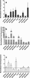
Differentially expressed tick selenogenes in R. parkeri-infected (Rp+) adult female ticks on day 5 after feeding. The Rp+ and R. parkeri-free adult female ticks that fed on sheep (in Fig. 1) were removed from the host on day 5 post-infestation and then dissected for tissue collection (midguts, salivary glands, and ovaries). Tick midguts and salivary glands isolated from single ticks and ovarian tissues were pooled from five individually-dissected Rp+ and Rp ticks. Quantitative reverse transcriptase PCR (qRT-PCR) was used to determine the transcriptional expression levels of the tick selenogenes. Differential gene expression was estimated in a tick midguts, b salivary glands, and c ovarian tissues. The expression levels in the Rp tick tissues were set to 1, as represented by dashed lines. eEFSec: selenocysteine elongation factor, SELENOM: selenoprotein M, SELENOK: selenoprotein K, SELENOS: selenoprotein S, SELENOO: selenoprotein O, TrxR: thioredoxin reductase, GST: glutathione S-transferase, SELENON: selenoprotein N, SELENOX: selenoprotein X, SELENOT: selenoprotein T
Fig. 5
Fig. 5
Functional characterization of tick selenoprotein gene knockdowns in R. parkeri-infected (Rp+) A. maculatum. A dsRNA-based silencing assay was performed for a SELENOM, b SELENOO, and c SELENOS in Rp+ ticks, and the compensatory expression levels of tick antioxidants and selenoproteins were estimated. The dsRNA specific for each selenogene (SELENOM, SELENOO, and SELENOS) was synthesized to include the addition of a T7 RNA polymerase binding site as the flanking sequence in the individual selenogene PCR amplicons from the dsRNA (Additional file 5: Table S1) and the in vitro RNA synthesis (which utilized the HiScribe™ T7 High Yield RNA synthesis kit, New England Biolabs). The dsRNA synthesized for each selenogene, along with irrelevant dsLacZ, were microinjected into 25–30 Rp+ ticks or 25–30 R. parkeri-free ticks (Additional file 3: Figure S3). The microinjected ticks were allowed to replete on sheep, and 5–10 ticks were removed from them to study the impact on gene silencing and the impact on Rickettsia parkeri and other symbionts (Figs. 6 and 7) on day 5 post-infestation. In each selenogene-silenced tick tissue (a SELENOM, b SELENOO, and c SELENOS), the transcript levels of a panel of selenogenes (SELENOM, SELENOO, and SELENOS, along with eEFSec, TrxR, SELENOK, SELENON, SELENOT) and redox genes (Cu/Zn-SOD, Mn-SOD, Duox, Catalase, GSHR, Salp25D) were measured. The transcript level for each gene in the control tissues was normalized to 1 for reference and is represented here as a dashed line. Tick GAPDH was used as a reference gene for normalizing the qRT-PCR results. d Oxidative stress in the selenogene-silenced tick midguts and control (dsLacZ) midguts was estimated using a malondialdehyde assay. KD, knockdown
Fig. 6
Fig. 6
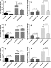
The impact of tick selenoprotein gene silencing on total bacterial and R. parkeri loads in the tick. The selenogene-silenced tick tissues (from Fig. 5) 5 days after microinjections of dsRNA–SELENOM, dsRNA–SELENOO, or dsRNA–SELENOS were used to estimate the total bacterial load (BL) and R. parkeri (Rp) load in the SELENOM- (a, b), SELENOO- (c, d), and SELENOS-silenced ticks (e, f). The qRT-PCR assay described in Figs. 1 and 2 was used to estimate the total bacterial load and Rp load per tick GAPDH. The p value is provided to compare statistical significance between the selenogene-silenced ticks and the control ticks. A p value of < 0.05 was considered statistically significant
Fig. 7
Fig. 7
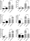
The impact of selenoprotein silencing on tick symbionts in R. parkeri (Rp+)-infected A. maculatum. The selenogene-silenced tick tissues (from Fig. 5) on day 5 post-microinjection of dsRNA–SELENOM, dsRNA–SELENOO, or dsRNA–SELENOS were used to determine the total bacterial load (BL) and Rp load in SELENOM- (a, b), SELENOO- (c, d), and SELENOS-silenced ticks (e, f). The qRT-PCR assay described in Figs. 1 and 2 was used to estimate the total bacterial load and Rp load per tick GAPDH. The p value is provided to compare the statistical significance between the selenogene-silenced ticks and the control ticks. A p value of < 0.05 was considered statistically significant
Fig. 8
Fig. 8
Proposed model for Rickettsia–symbiont interactions and the functional significance of selenoproteins in rickettsial replication inside tick tissues. The colonization of R. parkeri in A. maculatum gives it an opportunity to dynamically interact with tick symbionts and modulate tick defenses by regulating tick gene expression (e.g., selenogenes). In R. parkeri-free (Rp) tick cells, FLE and CMM are present and normal expression levels of tick selenoproteins (SELENOM, SELENOO, and SELENOS) occur. In R. parkeri-infected (Rp+) tick cells, the normal symbiont dynamics are altered such that CMM replicates and FLE numbers decline, and the selenoprotein expression levels are upregulated. Knocking down the selenoproteins by RNA interference reduces the selenoprotein expression levels, and the elevated levels of reactive oxygen species impair the replication of both R. parkeri and CMM while FLE replicates at normal levels. SELENOM (SELM)-SELENO (SELO)-SELENOS (SELS)

Similar articles

Cited by

References

    1. Raoult D, Roux V. Rickettsioses as paradigms of new or emerging infectious diseases. Clin Microbiol Rev. 1997;10:694–719. - PMC - PubMed
    1. Paddock CD, Goddard J. The evolving medical and veterinary importance of the Gulf Coast tick (Acari: Ixodidae) J Med Entomol. 2015;52:230–252. doi: 10.1093/jme/tju022. - DOI - PubMed
    1. Walker DH, Ismail N. Emerging and re-emerging rickettsioses: endothelial cell infection and early disease events. Nat Rev Microbiol. 2008;6:375–386. doi: 10.1038/nrmicro1866. - DOI - PubMed
    1. Parola P, Paddock CD, Raoult D. Tick-borne rickettsioses around the world: emerging diseases challenging old concepts. Clin Microbiol Rev. 2005;18:719–756. doi: 10.1128/CMR.18.4.719-756.2005. - DOI - PMC - PubMed
    1. Rikihisa Y. Anaplasma phagocytophilum and Ehrlichia chaffeensis: subversive manipulators of host cells. Nat Rev Microbiol. 2010;8:328–339. doi: 10.1038/nrmicro2318. - DOI - PubMed

Publication types

MeSH terms

LinkOut - more resources