. 2018 Aug:135:128-136.
doi: 10.1016/j.plefa.2018.07.015. Epub 2018 Jul 23.

Concentrations of oxidized linoleic acid derived lipid mediators in the amygdala and periaqueductal grey are reduced in a mouse model of chronic inflammatory pain

Affiliations

Concentrations of oxidized linoleic acid derived lipid mediators in the amygdala and periaqueductal grey are reduced in a mouse model of chronic inflammatory pain

J R Jensen et al. Prostaglandins Leukot Essent Fatty Acids. 2018 Aug.

Abstract

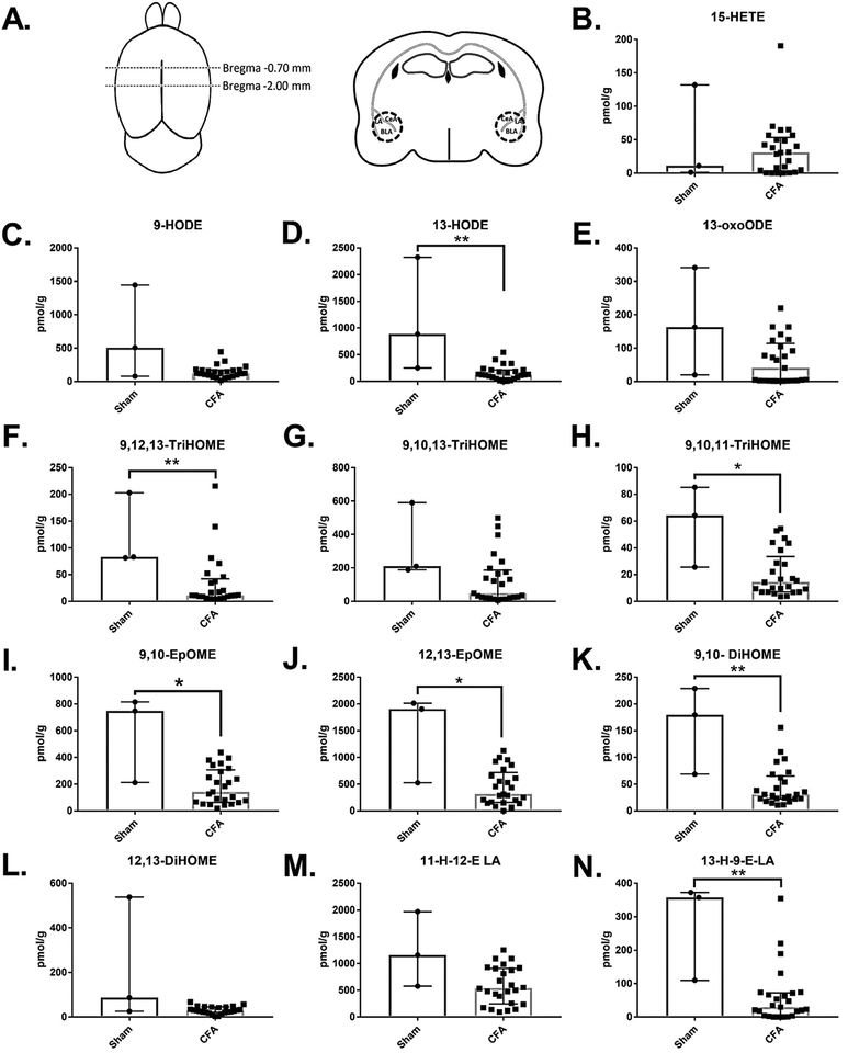
Chronic pain is both a global public health concern and a serious source of personal suffering for which current treatments have limited efficacy. Recently, oxylipins derived from linoleic acid (LA), the most abundantly consumed polyunsaturated fatty acid in the modern diet, have been implicated as mediators of pain in the periphery and spinal cord. However, oxidized linoleic acid derived mediators (OXLAMs) remain understudied in the brain, particularly during pain states. In this study, we employed a mouse model of chronic inflammatory pain followed by a targeted lipidomic analysis of the animals' amygdala and periaqueductal grey (PAG) using LC-MS/MS to investigate the effect of chronic inflammatory pain on oxylipin concentrations in these two brain nuclei known to participate in pain sensation and perception. From punch biopsies of these brain nuclei, we detected twelve OXLAMs in both the PAG and amygdala and one arachidonic acid derived mediator, 15-HETE, in the amygdala only. In the amygdala, we observed an overall decrease in the concentration of the majority of OXLAMs detected, while in the PAG the concentrations of only the epoxide LA derived mediators, 9,10-EpOME and 12,13-EpOME, and one trihydroxy LA derived mediator, 9,10,11-TriHOME, were reduced. This data provides the first evidence that OXLAM concentrations in the brain are affected by chronic pain, suggesting that OXLAMs may be relevant to pain signaling and adaptation to chronic pain in pain circuits in the brain and that the current view of OXLAMs in nociception derived from studies in the periphery is incomplete.


Figures

Fig. 1.
Fig. 1.. Chronic inflammatory pain reduced the concentration of most OXLAMs quantified in punch biopsies of the amygdala.
A. Schematic of landmarks used during tissue collection. The brain was blocked between approximately Bregma −0.70 mm and Bregma −2.00 mm, and a 1 mm circular punch biopsy, the location of which is demarcated by the broken line, was collected bilaterally including the majority of the BLA and portions of the LA and CeA. B-N. Concentrations of AA derived (B) and LA derived (C-N) oxylipins measured in amygdala punch biopsies using LC-MS/MS reveal a statistically significant reduction in most (D, F, H-K,N) but not all oxylipins in animals experiencing chronic inflammatory pain (n = 25) compared to sham treated controls (n = 3). Individual values are displayed as pmol per gram of tissue. Bars represent median (interquartile range). P-values were determined using the Mann-Whitney U test. *p < 0.05 and **p < 0.01 when comparing tissue collected from sham treated and CFA treated animals. BLA, basolateral amygdala; LA, lateral amygdala; CeA, central nucleus of the amygdala; HETE, hydroxyeicosatetraenoic acid; HODE, hydroxyoctadecadienoic acid; oxoODE, oxooctadecadienoic acid; TriHOME, trihydroxyoctadecenoic acid; EpOME, epoxyoctadecenoic acid; DiHOME, dihydroxyoctadecenoic acid; 11-H-12-E LA,11‑hydroxy‑12,13-trans-epoxy-octadecenoic acid; 13-H-9-E LA, 13‑hydroxy‑9,10-trans-epoxy-octadecenoic acid.
Fig. 2.
Fig. 2.. Chronicinflammatory pain reduced the concentration of some OXLAMs quantified in punch biopsies of the PAG.
A. Schematic of landmarks used during tissue collection. The brain was blocked between approximately Bregma −4.00 mm and Bregma −5.00 mm, and a 1 mm circular punch biopsy, the location of which is demarcated by the broken line, was taken using visual landmarks including the majority of the PAG. B-M. Concentration of OXLAMs measured in PAG punch biopsies using LC-MS/MS revealed a significant reduction in some (G-I) but not all OXLAMs in animals experiencing chronic inflammatory pain (n = 19) compared to sham treated controls (n = 3). Individual values are displayed as pmol per gram of tissue. Bars represent median (interquartile range). P-values were determined using the Mann-Whitney U test. *p < 0.05 and **p < 0.01 when comparing tissue collected from sham treated and CFA treated animals. PAG, periaqueductal grey; HODE, hydroxyoctadecadienoic acid; oxoODE, oxooctadecadienoic acid; TriHOME, trihydroxyoctadecenoic acid; EpOME, epoxyoctadecenoic acid; DiHOME, dihydroxyoctadecenoic acid; 11-H-12-E LA, 11‑hydroxy, 11‑hydroxy‑12,13-trans-epoxy-octadecenoic acid; 13-H-9-E LA, 13‑hydroxy‑9,10-trans-epoxyoctadecenoic acid.
Fig. 3.
Fig. 3.. Diagram of oxylipin synthesis from linoleic acid summarizing current findings.
Three weeks of chronic inflammatory pain affected the concentration of OXLAMs in multiple synthetic pathways in the amygdala, with the largest reduction in the concentration of 13-HODE relative to the mean concentration in sham treated controls. The oxylipin lowering effect of the chronic inflammatory pain model was limited to the epoxygenation products of LA in the PAG, with the exception of 9,10,11-TriHOME which was also the most reduced relative to the sham treated controls. OXLAM species shaded light grey highlight species with lower concentrations in the amygdala of CFA treated animals, while species shaded dark grey highlight species with lower concentrations in both the amygdala and PAG in this group. HETE, hydroxyeicosatetraenoic acid; HODE, hydroxyoctadecadienoic acid; oxoODE, oxooctadecadienoic acid; TriHOME, trihydroxyoctadecenoic acid; EpOME, epoxyoctadecenoic acid; DiHOME, dihydroxyoctadecenoic acid; 11-H-12-E LA,11‑hydroxy‑12,13-trans-epoxy-octadecenoic acid; 13-H-9-E LA, 13‑hydroxy‑9,10-trans-epoxy-octadecenoic acid.

References

    1. Bazinet RP, Layé S, Polyunsaturated fatty acids and their metabolites in brain function and disease, Nat. Rev. Neurosci 15 (2014) 771–785, https://doi.org/10.1038/nrn3820. - DOI - PubMed
    1. Chen CT, Green JT, Orr SK, Bazinet RP, Regulation of brain polyunsaturated fatty acid uptake and turnover, Prostaglandins Leukot Essent Fatty Acids 79 (2008) 85–91, https://doi.org/10.1016/j.plefa.2008.09.003. - DOI - PubMed
    1. Farooqui AA, Horrocks LA, Phospholipase A2-generated lipid mediators in the brain: the good, the bad, and the ugly, Neuroscientist 12 (2006) 245–260, https://doi.org/10.1177/1073858405285923. - DOI - PubMed
    1. Chang C-Y, Ke D-S, Chen J-Y, Essential fatty acids and human brain, Acta Neurol. Taiwan 18 (2009) 231–241. - PubMed
    1. Haag M, Essential fatty acids and the brain, Can. J. Psychiatry 48 (2003) 195–203, https://doi.org/10.1177/070674370304800308. - DOI - PubMed

Publication types