. 2018 Oct 25;86(11):e00397-18.
doi: 10.1128/IAI.00397-18. Print 2018 Nov.

In Vivo Virulence Characterization of Pregnancy-Associated Listeria monocytogenes Infections

Affiliations

In Vivo Virulence Characterization of Pregnancy-Associated Listeria monocytogenes Infections

Holly A Morrison et al. Infect Immun. .

Abstract

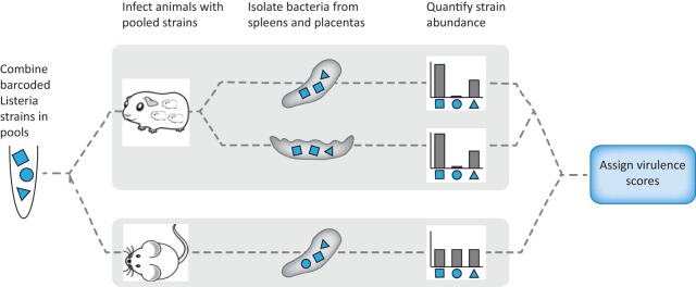
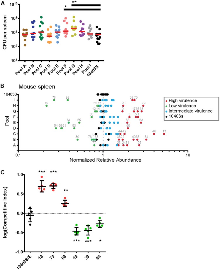
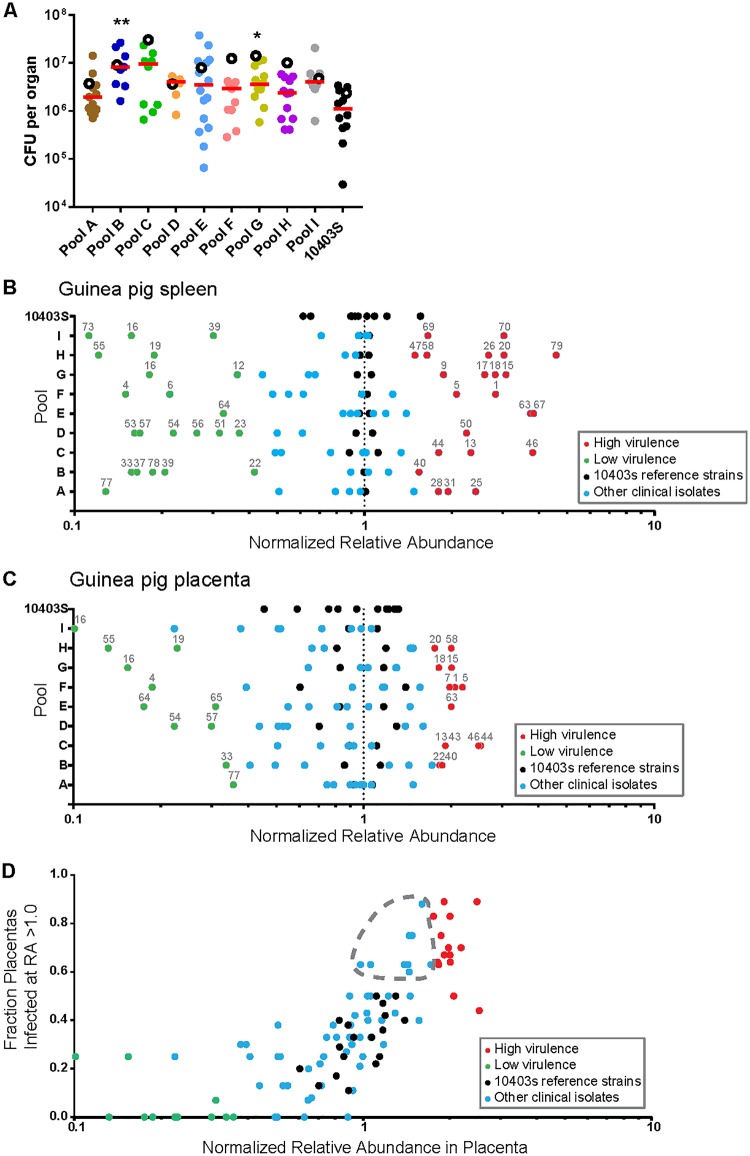
Listeria monocytogenes is a foodborne pathogen that infects the placenta and can cause pregnancy complications. Listeriosis usually occurs as a sporadic infection, but large outbreaks are also reported. Virulence from clinical isolates is rarely analyzed due to the large number of animals required, but this knowledge could help guide the response to an outbreak. We implemented a DNA barcode system using signature tags that allowed us to efficiently assay variations in virulence across a large number of isolates. We tested 77 signature-tagged clones of clinical L. monocytogenes strains from 72 infected human placentas and 5 immunocompromised patients, all of which were isolated since 2000. These strains were tested for virulence in a modified competition assay in comparison to that of the laboratory strain 10403S. We used two in vivo models of listeriosis: the nonpregnant mouse and the pregnant guinea pig. Strains that were frequently found at a high abundance within infected organs were considered hypervirulent, while strains frequently found at a low abundance were considered hypovirulent. Virulence split relatively evenly among hypovirulent strains, hypervirulent strains, and strains as virulent as 10403S. The laboratory strain was found to have an intermediate virulence phenotype, supporting its suitability for use in pathogenesis studies. Further, we found that splenic virulence and placental virulence are closely linked in both the guinea pig and mouse models. This suggests that outbreak and sporadic pregnancy-associated L. monocytogenes strains are not generally more virulent than lab reference strains. However, some strains did show consistent and reproducible virulence differences, suggesting that their further study may reveal deeper insights into the biological underpinnings of listeriosis.

Keywords: DNA barcode; Listeria; clinical isolate; epidemiology; placental infection; placental pathogen; signature tag; virulence.


Figures

FIG 1
FIG 1
Experimental design. Signature-tagged L. monocytogenes strains were pooled and injected i.v. into pregnant guinea pigs or nonpregnant mice. Each pool contained 11 barcoded strains: 9 clinical and 2 laboratory reference strains (10403S) in the clinical strain pools and 11 laboratory reference strains in the strain 10403S pool. For each organ set (guinea pig spleen, guinea pig placenta, mouse spleen), virulence scores were assigned to each strain on the basis of the average relative abundance in the infected organs in comparison to that of the laboratory reference strains.
FIG 2
FIG 2
Clinical isolates. (A) Pregnancy-associated L. monocytogenes strains (n = 72) from 25 U.S. states were collected by the CDC between 2000 and 2010, and 5 strains were isolated from immunocompromised patients at MSKCC (n = 5; immunocompromised). Most of the pregnancy-associated strains were associated with sporadic cases of listeriosis and were isolated from placental tissue (n = 68; pregnancy, sporadic). Four strains were associated with listeriosis outbreaks in the United States (n = 4; pregnancy, outbreak). These 4 strains were isolated from placenta (n = 2), maternal blood (n = 1), and neonatal blood (n = 1). (B) Serotype distribution of pregnancy-associated strains.
FIG 3
FIG 3
Virulence screen of clinical L. monocytogenes isolates in murine spleen. CD1 mice (nonpregnant) were infected i.v. with bacterial pools containing differentially tagged L. monocytogenes strains at equal ratios (total of 10 pools). Pools A to I contained 9 clinical strains and 2 10403S strains per pool; the 10403S pool contained 11 laboratory reference strains. Statistically significant differences in splenic bacterial burden from those in the control group were determined using one-way ANOVA with Dunnett's multiple comparisons posttest. ***, P < 0.0001; **, P < 0.01; *, P < 0.05. (A) Bacterial burden in murine spleen at 48 hpi with 2 × 10⁵ CFU per pool. For pools A to I there were 10 mice per pool; the 10403S pool contained 15 mice. Each circle represents the bacterial burden in one spleen, and each pool is represented by a different color. Red lines represent medians. (B) The average relative abundance of each strain in mouse spleen was quantified by qPCR. To accurately compare values across pools, the average relative abundance for each isolate was then normalized to the average for the reference strain in each pool. Significance Z-scores were calculated for the deviation from the range expected on the basis of the results for the 10403S pool (black circles). Blue circles indicate isolates with virulence similar to that of 10403S (intermediate virulence). Red and green circles indicate isolates with significantly higher and lower virulences, respectively. (C) CD1 mice were infected with one erythromycin-resistant 10403S strain and one erythromycin-susceptible untagged clinical isolate at a 1:1 ratio. The clinical isolates were chosen on the basis of their virulence scores in panel B: 3 hypervirulent (red circles) and 3 hypovirulent (green circles) strains. Competitive indices (isolate/10403S) were calculated for bacteria recovered from the spleen at 48 hpi. The control group was infected with two 10403S strains that differed in their susceptibility to erythromycin (10403S/E; black circles). Each group contained 5 mice from 2 separate experiments.
FIG 4
FIG 4
Virulence screen of clinical L. monocytogenes isolates in pregnant guinea pigs (spleen and placenta). Pregnant Hartley guinea pigs were infected i.v. with pools containing differentially tagged L. monocytogenes strains (Fig. 3). Statistically significant differences in the bacterial burden in the spleen and placenta from those in the control group were determined using one-way ANOVA with Dunnett's multiple comparisons posttest. **, P < 0.01; *, P < 0.05. (A) Bacterial burden in guinea pig spleen and placenta at 24 hpi with 108 CFU per pool. The total number of guinea pigs was 27 with a total of 107 placentas. The number of placentas in each pool was as follows: pool A, 12; pool B, 8; pool C, 9; pool D, 8; pool E, 15; pool F, 10; pool G, 14; pool H, 12; pool I, 8; strain 10403S pool, 11. Each filled circle represents the bacterial burden in one placenta, and each pool is represented by a different color. Red lines represent the median number of placental CFU. Empty circles represent the median bacterial burden in spleens from each pool. (B) The average relative abundance of each strain in guinea pig spleen was quantified by qPCR, and significance Z-scores were calculated. Black dots indicate 10403S strains. Blue circles indicate isolates with virulence similar to that of 10403S (intermediate virulence). Red and green circles indicate isolates with significantly higher and lower virulences, respectively. (C) Average relative abundance of each strain in guinea pig placenta quantified and calculated as described above. (D) Correlation of the relative abundance of each strain in the placenta with the fraction of placentas that it infected at a relative abundance higher than that of its inoculant (RA > 1.0). The gray dashed outline encircles isolates that were not identified to be highly virulent by relative abundance alone but for which infected fractions comparable to those for high-virulence isolates. The color coding corresponds to that in panel C.

Similar articles

Cited by

References

    1. Chenal-Francisque V, Lopez J, Cantinelli T, Caro V, Tran C, Leclercq A, Lecuit M, Brisse S. 2011. Worldwide distribution of major clones of Listeria monocytogenes. Emerg Infect Dis 17:1110–1112. doi:10.3201/eid/1706.101778. - DOI - PMC - PubMed
    1. de Noordhout CM, Devleesschauwer B, Angulo FJ, Verbeke G, Haagsma J, Kirk M, Havelaar A, Speybroeck N. 2014. The global burden of listeriosis: a systematic review and meta-analysis. Lancet Infect Dis 14:1073–1082. doi:10.1016/S1473-3099(14)70870-9. - DOI - PMC - PubMed
    1. Scallan E, Hoekstra RM, Angulo FJ, Tauxe RV, Widdowson M-A, Roy SL, Jones JL, Griffin PM. 2011. Foodborne illness acquired in the United States—major pathogens. Emerg Infect Dis 17:7–15. doi:10.3201/eid1701.P11101. - DOI - PMC - PubMed
    1. Lorber B. 1997. Listeriosis. Clin Infect Dis 24:1–9. - PubMed
    1. McCollum JT, Cronquist AB, Silk BJ, Jackson KA, O'Connor KA, Cosgrove S, Gossack JP, Parachini SS, Jain NS, Ettestad P, Ibraheem M, Cantu V, Joshi M, DuVernoy T, Fogg NW Jr, Gorny JR, Mogen KM, Spires C, Teitell P, Joseph LA, Tarr CL, Imanishi M, Neil KP, Tauxe RV, Mahon BE. 2013. Multistate outbreak of listeriosis associated with cantaloupe. N Engl J Med 369:944–953. doi:10.1056/NEJMoa1215837. - DOI - PubMed

Publication types

MeSH terms

Substances

LinkOut - more resources