Review
. 2018 Oct 11;132(15):1553-1560.
doi: 10.1182/blood-2018-03-784116. Epub 2018 Aug 13.

Chronic immune response dysregulation in MDS pathogenesis

Affiliations
Review

Chronic immune response dysregulation in MDS pathogenesis

Laura Barreyro et al. Blood. .

Abstract

Chronic innate immune signaling in hematopoietic cells is widely described in myelodysplastic syndromes (MDS), and innate immune pathway activation, predominantly via pattern recognition receptors, increases the risk of developing MDS. An inflammatory component to MDS has been reported for many years, but only recently has evidence supported a more direct role of chronic innate immune signaling and associated inflammatory pathways in the pathogenesis of MDS. Here we review recent findings and discuss relevant questions related to chronic immune response dysregulation in MDS.


Conflict of interest statement

Conflict-of-interest disclosure: The authors declare no competing financial interests.

Figures

None
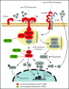
Graphical abstract
Figure 1.
Figure 1.
Cell intrinsic dysregulation of innate immune signaling in MDS HSCs. TLRs and interleukin-1 receptor (IL-1R)/IL1RAP recruit MyD88 and IRAK4/2 (Myddosome complex) upon ligand binding (lipopolysaccharide [LPS], S100A alarmins, and IL-1). CD14 functions as a coreceptor of TLR4 in response to LPS. Toll-interleukin 1 receptor (TIR) domain containing adaptor protein (TIRAP) can also increase the efficiency of Myddosome assembly by binding MyD88. IRAK4, a serine/threonine kinase, activates IRAK2 and/or IRAK1 through IRAK4-dependent phosphorylation. IRAK1 activates the ubiquitin (Ub) ligase, TRAF6, which mediates signaling to NF-κB, MAPK, and RNA binding proteins (ie, hnRNPA1) through K63-linked Ub chains, leading to expression of proinflammatory cytokines and NLRP3 or splicing of the Rho guanosine triphosphatase–activating protein Arhgap. microRNA-146a (miR-146a) suppresses IRAK1 and TRAF6 protein expression. miR-145 suppresses TIRAP protein expression. TIFAB suppresses TRAF6 protein stability. Inflammosome activation results in caspase 1–dependent IL-1β processing and pyroptosis. Proteins and genes in green are downregulated and/or deleted in MDS. Proteins and genes in red are overexpressed and/or activated in MDS. Steps of the signaling pathway that have been pharmacologically inhibited are indicated. Adapted from Varney et al.
Figure 2.
Figure 2.
Model of innate immune signaling dysregulation in the pathogenesis of MDS. Certain diseases and conditions, such as aging, autoimmune disorders, chronic infections, and/or clonal hematopoiesis of indeterminate potential (CHIP), can induce innate immune signaling dysregulation in HSCs in part by creating an inflammatory BM microenvironment characterized by increased alarmins and/or cytokines. Development of MDS may occur by at least 2 independent mechanisms. (1) CHIP-associated mutations (ie, DNMT3a or TET2) occur in HSCs by innate immune independent mechanisms and drive the expansion of myeloid-biased HSC leading to altered innate immune signaling and development of MDS. (2) Prolonged innate immune signaling caused by clonally expanded myeloid-biased HSCs directly increases the risk of acquiring mutations (ie, CHIP mutations) contributing to MDS. Innate immune signaling dysregulation at the MDS stage occurs through cell-intrinsic (ie, increased cell death via pyroptosis) and cell-extrinsic mechanisms (ie, cytokines and alarmins stimulation from macrophage and myeloid derived suppressor cells [MDSCs]). As a result of altered innate immune signaling, MDSCs also promote regulatory T cell (Treg) activation to limit T-cell surveillance.

References

    1. Esplin BL, Shimazu T, Welner RS, et al. . Chronic exposure to a TLR ligand injures hematopoietic stem cells. J Immunol. 2011;186(9):5367-5375. - PMC - PubMed
    1. Hofmann WK, de Vos S, Komor M, Hoelzer D, Wachsman W, Koeffler P. Characterization of gene expression of CD34+ cells from normal and myelodysplastic bone marrow. Blood. 2002;100(10):3553-3560. - PubMed
    1. Pellagatti A, Cazzola M, Giagounidis A, et al. . Deregulated gene expression pathways in myelodysplastic syndrome hematopoietic stem cells. Leukemia. 2010;24:756-764. - PubMed
    1. Maratheftis CI, Andreakos E, Moutsopoulos HM, Voulgarelis M. Toll-like receptor-4 is up-regulated in hematopoietic progenitor cells and contributes to increased apoptosis in myelodysplastic syndromes. Clin Cancer Res. 2007;13(4):1154-1160. - PubMed
    1. Wei Y, Dimicoli S, Bueso-Ramos C, et al. . Toll-like receptor alterations in myelodysplastic syndrome. Leukemia. 2013;27(9):1832-1840. - PMC - PubMed

Publication types

MeSH terms