. 2018 Oct 24;62(11):e01142-18.
doi: 10.1128/AAC.01142-18. Print 2018 Nov.

Disrupting Gram-Negative Bacterial Outer Membrane Biosynthesis through Inhibition of the Lipopolysaccharide Transporter MsbA

Affiliations

Disrupting Gram-Negative Bacterial Outer Membrane Biosynthesis through Inhibition of the Lipopolysaccharide Transporter MsbA

Mary Kate Alexander et al. Antimicrob Agents Chemother. .

Abstract

There is a critical need for new antibacterial strategies to counter the growing problem of antibiotic resistance. In Gram-negative bacteria, the outer membrane (OM) provides a protective barrier against antibiotics and other environmental insults. The outer leaflet of the outer membrane is primarily composed of lipopolysaccharide (LPS). Outer membrane biogenesis presents many potentially compelling drug targets as this pathway is absent in higher eukaryotes. Most proteins involved in LPS biosynthesis and transport are essential; however, few compounds have been identified that inhibit these proteins. The inner membrane ABC transporter MsbA carries out the first essential step in the trafficking of LPS to the outer membrane. We conducted a biochemical screen for inhibitors of MsbA and identified a series of quinoline compounds that kill Escherichia coli through inhibition of its ATPase and transport activity, with no loss of activity against clinical multidrug-resistant strains. Identification of these selective inhibitors indicates that MsbA is a viable target for new antibiotics, and the compounds we identified serve as useful tools to further probe the LPS transport pathway in Gram-negative bacteria.

Keywords: ABC transporters; Enterobacteriaceae; Escherichia coli; MsbA; lipopolysaccharide; outer membrane.


Figures

FIG 1
FIG 1
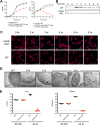
MsbA conditional deletion strain. (A) Growth of CFT073 wild type (WT) versus CFT073 msbA-cKO with or without arabinose (ara) to induce MsbA expression, as measured by OD600 (left) and CFU count (right). Data are shown from a representative experiment. (B) Western blots of MsbA protein expression over time. Loading was normalized by the OD600 value, and MsbA protein was detected using a rabbit polyclonal antibody generated in-house. (C) Confocal images of the CFT073 wild type versus the CFT073 msbA-cKO strain grown in the absence of arabinose for the indicated amount of time. Membrane is stained with Nile red, and DNA is stained with DAPI (blue). (D) TEM images of the CFT073 wild type versus the CFT073 msbA-cKO strain grown in the absence of arabinose for the indicated amount of time. Examples of vesicle-like membrane accumulation are indicated with black arrows, and parallel membrane stacks are indicated with white arrows. Lysed cells at the 4-h time point are indicated by open arrows. (E) CFT073 wild-type versus CFT073 msbA-cKO CFU counts isolated from mouse livers and spleens 30 min or 24 h following intravenous injection with 106 CFU in neutropenic mice. Ten mice were injected with each strain, and five were sacrificed at each time point. Two mice injected with the wild-type CFT073 died prior to the 24-h time point, so CFU counts are plotted for the remaining three mice.
FIG 2
FIG 2
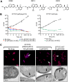
Small-molecule inhibitors of E. coli MsbA. (A) Chemical structures of the three quinoline inhibitors described in this paper. (B) Time-kill assay of CFT073 lptD(imp4213) and CFT073 with MsbA inhibitors. Viable cells were measured by plating on LB agar at 0, 1, 2, 3, 5, and 22 h after addition of compound. (C) Confocal microscopy and TEM of CFT073 and CFT073 lptD(imp4213) treated with the indicated concentration of inhibitor for 3 h. For confocal microscopy, membranes were stained with Nile red, and DNA was stained with DAPI (blue). Arrows in confocal and TEM images indicate sites of excess membrane accumulation. Untreated CFT073 cells looked identical to untreated CFT073 lptD(imp4213) cells (data not shown).
FIG 3
FIG 3
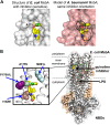
Binding site of MsbA inhibitors. (A) Comparison of inhibitor binding sites in E. coli MsbA (from crystal structure) and A. baumannii MsbA (modeled). (B) Location of inhibitor binding site in the full-length MsbA crystal structure (right) and location of quinoline-resistant mutations within the binding site. NBD, nucleotide binding domains.
FIG 4
FIG 4
E. coli stress response following treatment with MsbA inhibitors. (A) qRT-PCR of RNA isolated from E. coli CFT073 lptD(imp4213) treated for 2 h with 1.2 μM G913. Corresponding stress response pathways are indicated below gene names. (B) qRT-PCR of RNA isolated from CFT073 msbA-cKO grown in the absence of arabinose for 2 h or 4 h. (C) LPS gel showing colanic acid modification of LPS following MsbA depletion. WT, MG1655; msbA-cKO, MG1655 msbA-cKO; imp, MG1655 lptD(imp4213); RQ, relative quantification.

Similar articles

Cited by

References

    1. World Health Organization. 2014. Antimicrobial resistance: global report on surveillance. World Health Organization, Geneva, Switzerland: http://www.who.int/drugresistance/documents/surveillancereport/en/.
    1. Centers for Disease Control and Prevention. 2013. Antibiotic resistance threats in the United States, 2013. Centers for Disease Control and Prevention, Atlanta, GA: https://www.cdc.gov/drugresistance/threat-report-2013/pdf/ar-threats-201....
    1. Kamio Y, Nikaido H. 1976. Outer membrane of Salmonella typhimurium: accessibility of phospholipid head groups to phospholipase c and cyanogen bromide activated dextran in the external medium. Biochemistry 15:2561–2570. doi:10.1021/bi00657a012. - DOI - PubMed
    1. Osborn MJ, Gander JE, Parisi E. 1972. Mechanism of assembly of the outer membrane of Salmonella typhimurium. Site of synthesis of lipopolysaccharide. J Biol Chem 247:3973–3986. - PubMed
    1. Osborn MJ, Gander JE, Parisi E, Carson J. 1972. Mechanism of assembly of the outer membrane of Salmonella typhimurium. Isolation and characterization of cytoplasmic and outer membrane. J Biol Chem 247:3962–3972. - PubMed

Publication types

MeSH terms

LinkOut - more resources