. 2018 Oct 24;62(11):e00707-18.
doi: 10.1128/AAC.00707-18. Print 2018 Nov.

Nitazoxanide Inhibits Human Norovirus Replication and Synergizes with Ribavirin by Activation of Cellular Antiviral Response

Affiliations

Nitazoxanide Inhibits Human Norovirus Replication and Synergizes with Ribavirin by Activation of Cellular Antiviral Response

Wen Dang et al. Antimicrob Agents Chemother. .

Abstract

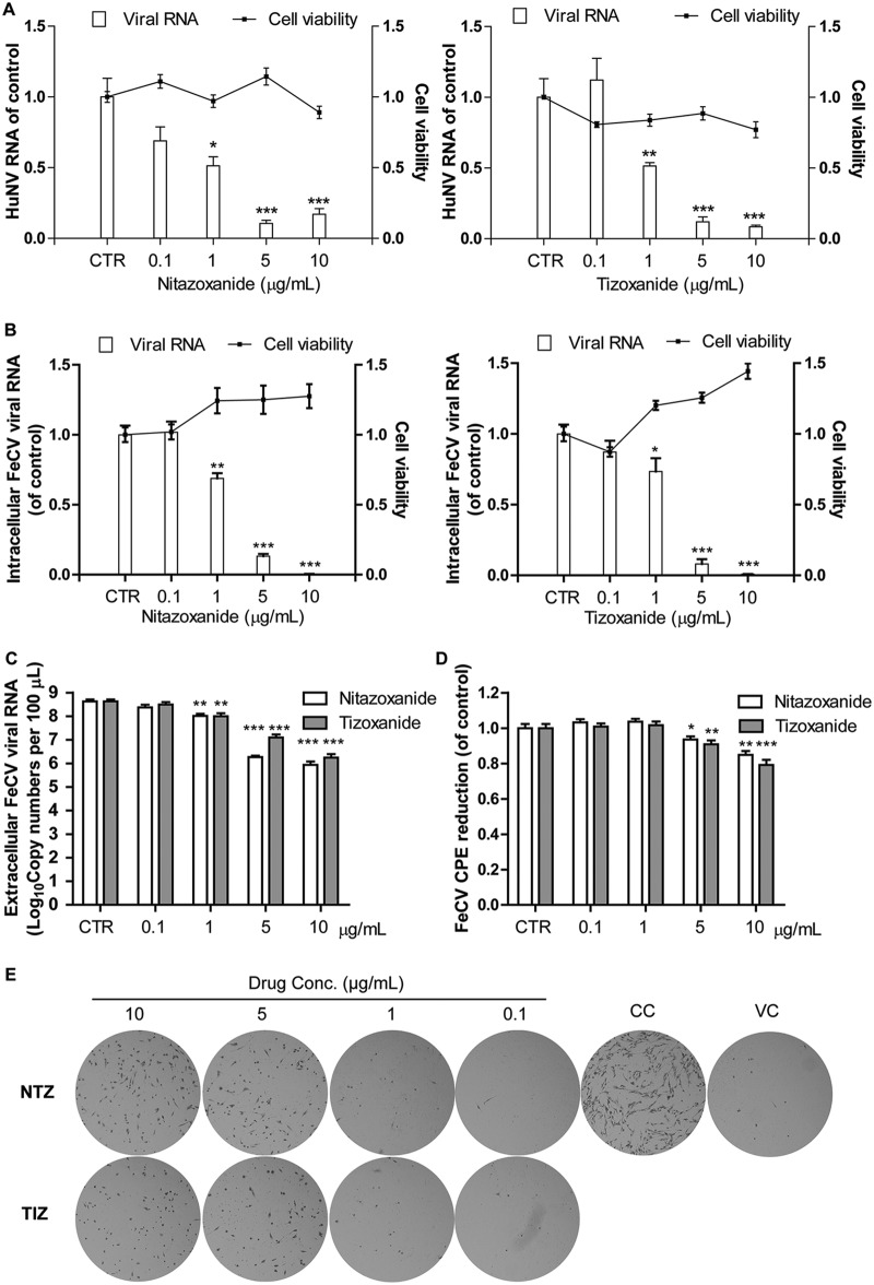
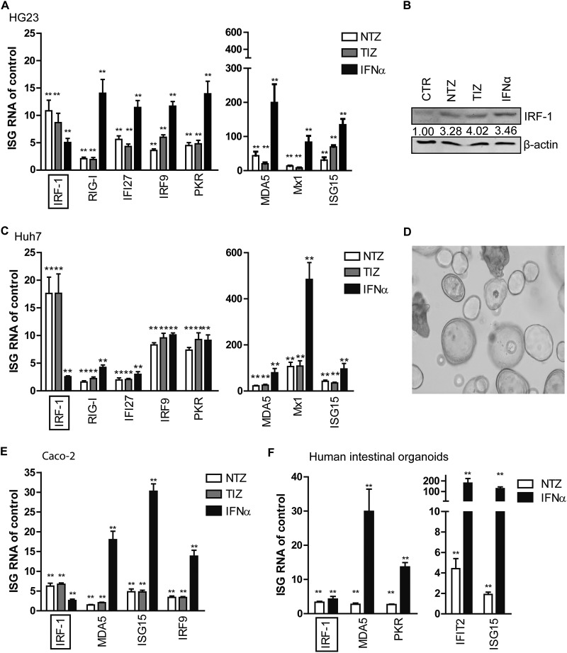
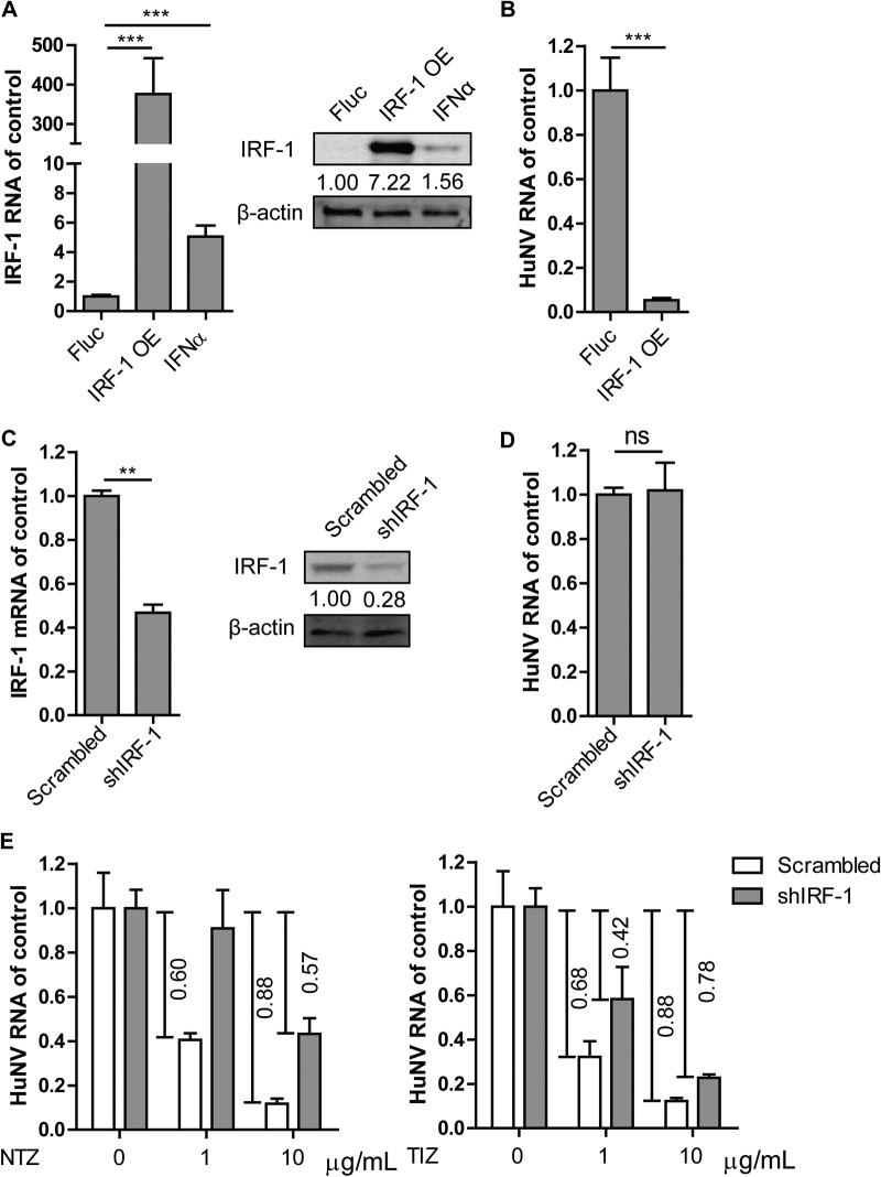
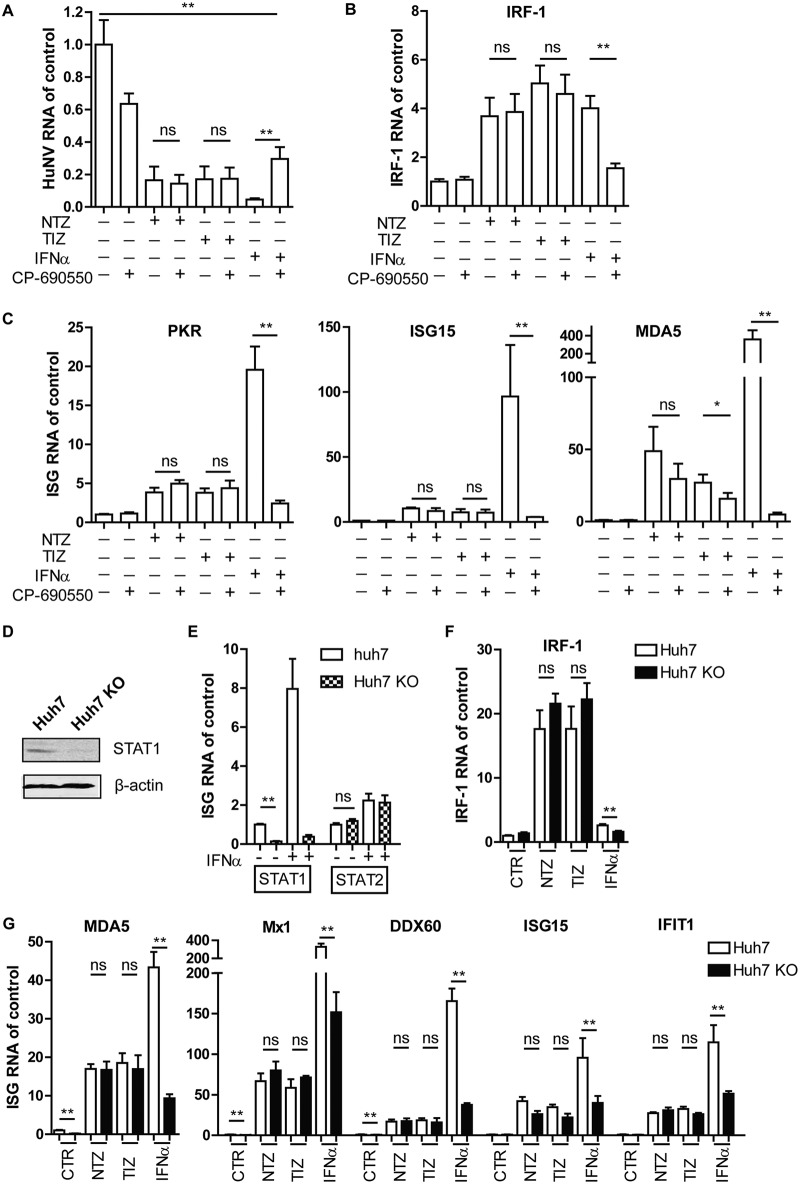
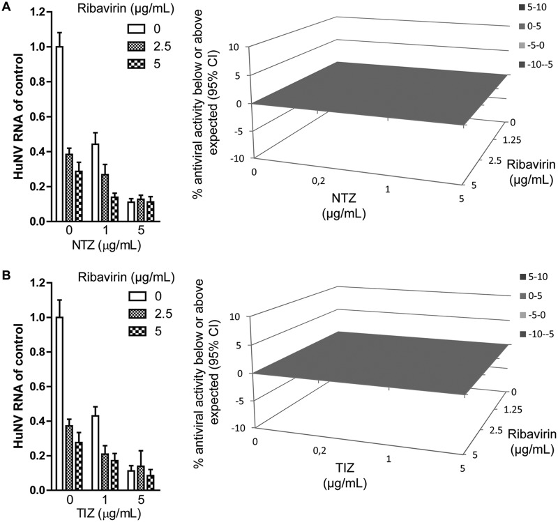
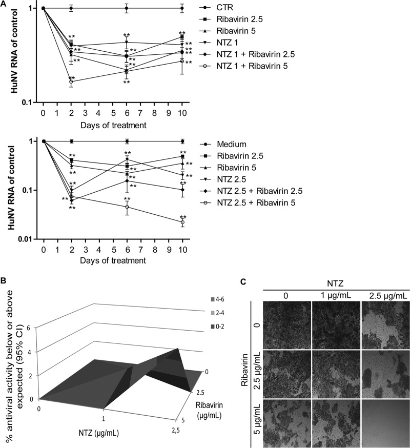
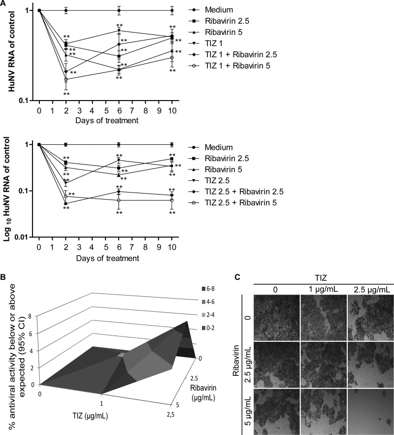
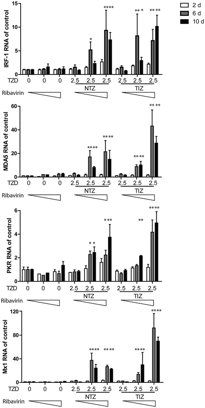
Norovirus is the main cause of viral gastroenteritis worldwide. Although norovirus gastroenteritis is self-limiting in immunocompetent individuals, chronic infections with debilitating and life-threatening complications occur in immunocompromised patients. Nitazoxanide (NTZ) has been used empirically in the clinic and has demonstrated effectiveness against norovirus gastroenteritis. In this study, we aimed at uncovering the antiviral potential and mechanisms of action of NTZ and its active metabolite, tizoxanide (TIZ), using a human norovirus (HuNV) replicon. NTZ and TIZ, collectively referred to as thiazolides (TZD), potently inhibited replication of HuNV and a norovirus surrogate, feline calicivirus. Mechanistic studies revealed that TZD activated cellular antiviral response and stimulated the expression of a subset of interferon-stimulated genes (ISGs), particularly interferon regulatory factor 1 (IRF-1), not only in a Huh7 cell-based HuNV replicon, but also in naive Huh7 and Caco-2 cells and novel human intestinal organoids. Overexpression of exogenous IRF-1 inhibited HuNV replication, whereas knockdown of IRF-1 largely attenuated the antiviral activity of TZD, suggesting that IRF-1 mediated TZD inhibition of HuNV. By using a Janus kinase (JAK) inhibitor, CP-690550, and a STAT1 knockout approach, we found that TZD induced antiviral response independently of the classical JAK-signal transducers and activators of transcription (JAK-STAT) pathway. Furthermore, TZD and ribavirin synergized to inhibit HuNV replication and completely depleted the replicons from host cells after long-term treatment. In summary, our results demonstrated that TZD combated HuNV replication through activation of cellular antiviral response, in particular by inducing a prominent antiviral effector, IRF-1. NTZ monotherapy or combination with ribavirin represent promising options for treating norovirus gastroenteritis, especially in immunocompromised patients.

Keywords: IRF-1; cell culture model; nitazoxanide; noroviruses; ribavirin; synergy; tizoxanide.


Figures

FIG 1
FIG 1
TZD potently inhibited replication of HuNV and its surrogate FeCV without significant cytotoxicity. (A) Nitazoxanide and its active metabolite tizoxanide dose-dependently inhibited HuNV replication without clear toxicity to host cells after 2 days of treatment. The level of HuNV replicon RNA was quantified using qRT-PCR and compared to that of vehicle control (0.05% DMSO, set as 1) (CTR) (n = 3 independent experiments, each in duplicate). (B) TZD elicited potent antiviral potential against FeCV. CRFK cells were first infected with FeCV (at an MOI of 0.1) and incubated with vehicle control or increasing concentrations of TZD. After 24 h of treatment, the cellular FeCV RNA level was quantified by qRT-PCR, normalized to that of feline GAPDH, and compared to vehicle control (set as 1). (C) Same as panel B for detecting cellular FeCV RNA; viral RNA copy numbers in the supernatant (secreted viruses) were also detected after 24 h of treatment with vehicle control or TZD. TZD significantly reduced extracellular FeCV RNA in the supernatant (n = 3 independent experiments, each in duplicate). (D and E) The anti-FeCV activity of TZD was further validated by an MTT- and hematoxylin and eosin staining-based CPE reduction assay. CRFK cells were first infected with FeCV (at an MOI of 0.5) and incubated with vehicle control or TZD. After 2 days of treatment, the CPE was quantified by MTT assay, and residual cells were observed after fixation and staining with hematoxylin and eosin. In panel E, CC and VC represent cell control and virus control, respectively. The images are representative of three independent experiments. The data are presented as means ± SEM (*, P < 0.05; **, P < 0.01; ***, P < 0.001).
FIG 2
FIG 2
TZD robustly stimulated the expression of IRF-1 and several other ISGs in a HuNV replicon model and intestinal models of Caco-2 cells and primary organoids. (A) Fold changes of ISGs induced by NTZ (10 μg/ml), TIZ (10 μg/ml), or IFN-α (1,000 IU/ml) in HG23 cells. After 2 days of treatment, the levels of ISG mRNA were quantified by qRT-PCR and normalized to those of human GAPDH. The results are expressed as fold changes compared to the vehicle control (DMSO; n = 3 independent experiments, each in duplicate). (B) Western blot analysis of IRF-1 protein expression in response to NTZ (10 μg/ml), TIZ (10 μg/ml), or IFN-α (1,000 IU/ml). The data are representative of the results of three independent experiments. (C) Naive Huh7 cells were treated with NTZ (10 μg/ml), TIZ (10 μg/ml), IFN-α (1,000 IU/ml), or vehicle control. After 2 days of treatment, relative levels of ISG mRNA were quantified by qRT-PCR (n = 3 independent experiments, each in duplicate). (D) Morphology of 3D human primary intestinal organoids in Matrigel. (E) Relative levels of ISG RNA were quantified by qRT-PCR after 2 days of treatment with NTZ (10 μg/ml) or IFN-α (1,000 IU/ml) in Caco-2 cells (n = 3 independent experiments, each in duplicate). (C) Relative levels of ISG RNA in human intestinal organoids after treatment with NTZ (10 μg/ml) or IFN-α (1,000 IU/ml) for 2 days (n = 3 independent experiments, each in duplicate). The data are presented as means and SEM (*, P < 0.05; **, P < 0.01).
FIG 3
FIG 3
IRF-1 mediated TZD-triggered inhibition of HuNV replication. (A) To overexpress (OE) IRF-1, HG23 cells were transduced with lentiviral IRF-1-expressing vector or Fluc-expressing vector (control). After 2 days of transduction, the levels of IRF-1 expression were detected by qRT-PCR (n = 3 independent experiments, each in duplicate) and Western blotting (n = 3 independent experiments). IFN-α (1,000 IU/ml) was used as a positive control. (B) Overexpression of IRF-1 potently inhibited HuNV replication (n = 3 independent experiments, each in duplicate). (C) To further evaluate the role of basal IRF-1 in HuNV replication, HG23 cells were transduced with lentiviral shRNA vector targeting IRF-1. A scrambled vector was used as a nontargeting control. Successful knockdown of IRF-1 was confirmed by qRT-PCR (n = 3 independent experiments, each in duplicate) and Western blotting (n = 3 independent experiments). (D) Knockdown of IRF-1 had minor effects on HuNV replication (n = 3 independent experiments, each in duplicate). (E) IRF-1 shRNA and nontargeting control cells were mock treated or treated with the indicated concentrations of TZD. After 2 days of treatment, the levels of HuNV RNA were quantified by qRT-PCR and compared to those of the respective control (n = 3 independent experiments, each in duplicate). The data are presented as means and SEM (**, P < 0.01; ***, P < 0.001; ns, not significant).
FIG 4
FIG 4
Stimulation of antiviral response by TZD was independent of the JAK-STAT pathway. (A) HG23 cells were treated with vehicle only (DMSO; control), NTZ (10 μg/ml), TIZ (10 μg/ml), or IFN-α (100 IU/ml) alone or in combination with CP-690550 (1,000 ng/ml). After 2 days of treatment, HuNV replication was quantified by qRT-PCR (n = 3 independent experiments, each in duplicate). (B and C) The expression of IRF-1 and other ISGs, including PKR, ISG15, and MDA5, was quantified by qRT-PCR (n = 3 independent experiments, each in duplicate). (D) A STAT1 KO clone was established from Huh7 cells expressing STAT1 sgRNAs. Successful KO of STAT1 was confirmed by Western blotting. (E) To further confirm successful KO of STAT1, Huh7 control and STAT1 KO cells were treated with IFN-α (1,000 IU/ml) for 2 days. The levels of STAT1 and STAT2 RNA were evaluated by qRT-PCR (n = 3 independent experiments, each in duplicate). KO of STAT1 abolished the induction of STAT1, but not STAT2, after IFN-α treatment. (F and G) Huh7 control and STAT1 KO cells were mock treated (DMSO; control) or treated with NTZ (10 μg/ml), TIZ (10 μg/ml), or IFN-α (1,000 IU/ml). After 2 days of treatment, the expression levels of IRF-1, MDA5, Mx1, DDX60, ISG15, and IFIT1 were detected by qRT-PCR. The results were first normalized to human GAPDH and then compared to control cells (n = 3 independent experiments, each in duplicate). The data are presented as means and SEM (*, P < 0.05; **, P < 0.01; ns, not significant).
FIG 5
FIG 5
TZD worked additively with ribavirin to inhibit HuNV replication after short-term treatment. (A) HG23 cells were treated with various concentrations of NTZ alone, ribavirin alone, or the two in combination for 2 days. (Left) Antiviral activities were determined by qRT-PCR (n = 3 independent experiments, each in duplicate). (Right) To further explore the drug-drug interaction, the antiviral results were analyzed with a mathematical model. The 3D surface plot shown represents the difference (within a 95% confidence interval [CI]) between actual experimental effects and the theoretical additive effect of the combination at various concentrations of the two compounds. (B) Combination of TIZ with ribavirin. The error bars represent SEM.
FIG 6
FIG 6
Long-term combination of NTZ with ribavirin synergistically inhibited HuNV replication and completely depleted HuNV replicons from host cells. (A) HG23 cells were incubated with NTZ alone, ribavirin alone, or the two drugs in combination. After 2 days of culture, the cells were passaged to fresh drug-containing medium for another 4 days of incubation. After 6 days, the cells were passaged with another round of 4 days of treatment. At the end of each treatment, the HG23 cells were harvested and analyzed for levels of HuNV RNA (n = 3 independent experiments, each in duplicate). (B) Synergy analysis. The antiviral results after 10 days of treatment were analyzed with MacSynergy. (C) Rebound assay. After 10 days of treatment, HG23 cells were plated into a 48-well plate (2 × 10⁵ cells per well) containing 250 μl medium with 1 mg/ml G418. After 5 days of culture, the cell colonies were stained and visualized using an inverted light microscope. The data presented are representative of 3 independent experiments. The data are presented as means ± SEM (**, P < 0.01).
FIG 7
FIG 7
Combining TIZ with ribavirin synergistically inhibited HuNV replication. (A) Combination of TIZ with ribavirin showed greater efficacy against HuNV replication than the individual drugs (n = 3 independent experiments, each in duplicate). (B) Synergy analysis. (C) Rebound assay after long-term treatment. The data are presented as means ± SEM (**, P < 0.01).
FIG 8
FIG 8
Augmented induction of ISGs after combination treatment with TZD and ribavirin. Shown are qRT-PCR analysis of expression levels for IRF-1, MDA5, and PKR after treatment with TZD (NTZ, 2.5 μg/ml; TIZ, 2.5 μg/ml) and ribavirin (0, 2.5, and 5 μg/ml) in HG23 cells for 2, 6, and 10 days (d) (n = 3 independent experiments, each in duplicate). The error bars indicate SEM.

Similar articles

Cited by

References

    1. Bartsch SM, Lopman BA, Ozawa S, Hall AJ, Lee BY. 2016. Global economic burden of norovirus gastroenteritis. PLoS One 11:e0151219. doi:10.1371/journal.pone.0151219. - DOI - PMC - PubMed
    1. Ye X, Van JN, Munoz FM, Revell PA, Kozinetz CA, Krance RA, Atmar RL, Estes MK, Koo HL. 2015. Noroviruses as a cause of diarrhea in immunocompromised pediatric hematopoietic stem cell and solid organ transplant recipients. Am J Transplant 15:1874–1881. doi:10.1111/ajt.13227. - DOI - PMC - PubMed
    1. Kaufman SS, Chatterjee NK, Fuschino ME, Morse DL, Morotti RA, Magid MS, Gondolesi GE, Florman SS, Fishbein TM. 2005. Characteristics of human calicivirus enteritis in intestinal transplant recipients. J Pediatr Gastroenterol Nutr 40:328–333. doi:10.1097/01.MPG.0000155182.54001.48. - DOI - PubMed
    1. Schwartz S, Vergoulidou M, Schreier E, Loddenkemper C, Reinwald M, Schmidt-Hieber M, Flegel WA, Thiel E, Schneider T. 2011. Norovirus gastroenteritis causes severe and lethal complications after chemotherapy and hematopoietic stem cell transplantation. Blood 117:5850–5856. doi:10.1182/blood-2010-12-325886. - DOI - PMC - PubMed
    1. Morris J, Morris C. 2015. Nitazoxanide is effective therapy for norovirus gastroenteritis after chemotherapy and hematopoietic stem cell transplantation (HSCT). Biol Blood Marrow Transplant 21:S255–S256.

Publication types

MeSH terms