

Spectroscopic ruler for measuring active-site distortions based on Raman optical activity of a hydrogen out-of-plane vibration
- PMID: 30104345
- PMCID: PMC6126711
- DOI: 10.1073/pnas.1806491115
Spectroscopic ruler for measuring active-site distortions based on Raman optical activity of a hydrogen out-of-plane vibration
Abstract
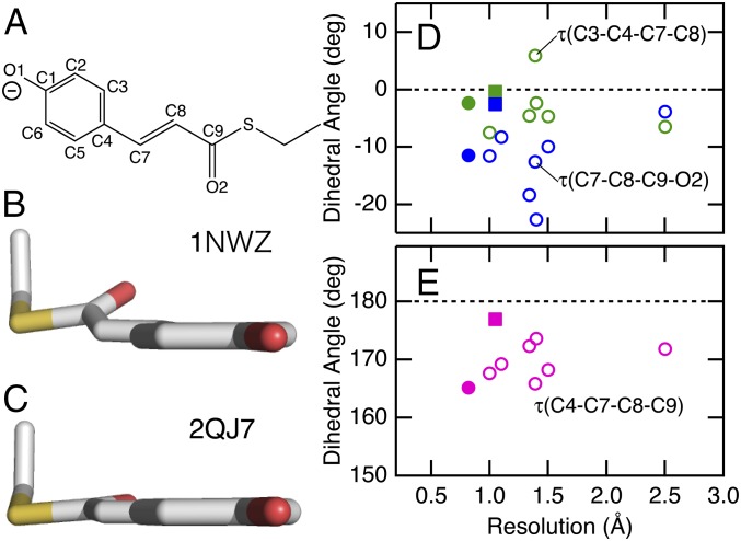
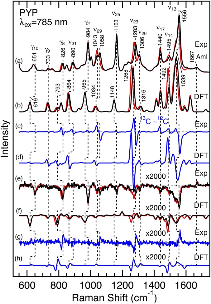
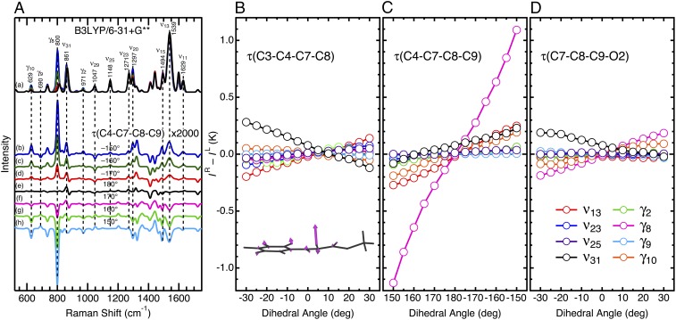
Photoactive yellow protein (PYP), from the phototrophic bacterium Halorhodospira halophila, is a small water-soluble photoreceptor protein and contains p-coumaric acid (pCA) as a chromophore. PYP has been an attractive model for studying the physical chemistry of protein active sites. Here, we explore how Raman optical activity (ROA) can be used to extract quantitative information on distortions of the pCA chromophore at the active site in PYP. We use 13C8-pCA to assign an intense signal at 826 cm-1 in the ROA spectrum of PYP to a hydrogen out-of-plane vibration of the ethylenic moiety of the chromophore. Quantum-chemical calculations based on density functional theory demonstrate that the sign of this ROA band reports the direction of the distortion in the dihedral angle about the ethylenic C=C bond, while its amplitude is proportional to the dihedral angle. These results document the ability of ROA to quantify structural deformations of a cofactor molecule embedded in a protein moiety.
Keywords: chromophore; density functional theory; molecular strain; photoreceptor; vibrational spectroscopy.
Conflict of interest statement
The authors declare no conflict of interest.
Figures
Similar articles
-
Resonance Raman spectroscopy and quantum chemical calculations reveal structural changes in the active site of photoactive yellow protein.Biochemistry. 2002 Apr 30;41(17):5668-74. doi: 10.1021/bi025508o. Biochemistry. 2002. PMID: 11969428
-
Vibrational assignment of the 4-hydroxycinnamyl chromophore in photoactive yellow protein.J Phys Chem B. 2007 Mar 15;111(10):2719-26. doi: 10.1021/jp066434j. Epub 2007 Feb 21. J Phys Chem B. 2007. PMID: 17311445
-
Photoinduced isomerization of the photoactive yellow protein (PYP) chromophore: interplay of two torsions, a HOOP mode and hydrogen bonding.J Phys Chem A. 2011 Aug 25;115(33):9237-48. doi: 10.1021/jp2011843. Epub 2011 Aug 3. J Phys Chem A. 2011. PMID: 21744877
-
Initial photoinduced dynamics of the photoactive yellow protein.Chemphyschem. 2005 May;6(5):828-37. doi: 10.1002/cphc.200400351. Chemphyschem. 2005. PMID: 15884065 Review.
-
Ultrafast spectroscopy of biological photoreceptors.Curr Opin Struct Biol. 2007 Oct;17(5):623-30. doi: 10.1016/j.sbi.2007.09.006. Epub 2007 Oct 23. Curr Opin Struct Biol. 2007. PMID: 17959372 Review.
Cited by
-
All-dielectric chiral-field-enhanced Raman optical activity.Nat Commun. 2021 May 24;12(1):3062. doi: 10.1038/s41467-021-23364-w. Nat Commun. 2021. PMID: 34031409 Free PMC article.
-
Induced Lanthanide Circularly Polarized Luminescence as a Probe of Protein Fibrils.ACS Omega. 2019 Jan 15;4(1):1265-1271. doi: 10.1021/acsomega.8b03175. eCollection 2019 Jan 31. ACS Omega. 2019. PMID: 31459399 Free PMC article.
-
Reisomerization of retinal represents a molecular switch mediating Na+ uptake and release by a bacterial sodium-pumping rhodopsin.J Biol Chem. 2022 Sep;298(9):102366. doi: 10.1016/j.jbc.2022.102366. Epub 2022 Aug 11. J Biol Chem. 2022. PMID: 35963435 Free PMC article.
-
Spectroscopic approach for exploring structure and function of photoreceptor proteins.Biophys Physicobiol. 2021 May 14;18:127-130. doi: 10.2142/biophysico.bppb-v18.014. eCollection 2021. Biophys Physicobiol. 2021. PMID: 34178563 Free PMC article. No abstract available.
-
Recognition of the True and False Resonance Raman Optical Activity.Angew Chem Int Ed Engl. 2021 Sep 20;60(39):21205-21210. doi: 10.1002/anie.202107600. Epub 2021 Aug 21. Angew Chem Int Ed Engl. 2021. PMID: 34216087 Free PMC article.
References
-
- Spudich JL, Yang C-S, Jung K-H, Spudich EN. Retinylidene proteins: Structures and functions from archaea to humans. Annu Rev Cell Dev Biol. 2000;16:365–392. - PubMed
-
- Boulanger E, Harvey JN. QM/MM methods for free energies and photochemistry. Curr Opin Struct Biol. 2018;49:72–76. - PubMed
-
- Rocha-Rinza T, Sneskov K, Christiansen O, Ryde U, Kongsted J. Unraveling the similarity of the photoabsorption of deprotonated p-coumaric acid in the gas phase and within the photoactive yellow protein. Phys Chem Chem Phys. 2011;13:1585–1589. - PubMed
Publication types
MeSH terms
Substances
LinkOut - more resources
Full Text Sources
Other Literature Sources
Miscellaneous
